Identifying Tissue of the Gastrointestinal Tract in Vivo
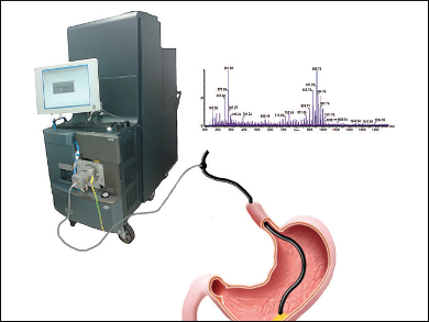
Cancer or no cancer? This question can usually only be answered after a days-long wait for a histological examination. With the use of a mass spectrometric technique, the answer may soon be available in real time. In the journal Angewandte Chemie, a team of British and Hungarian researchers has now introduced an endoscopic system that can differentiate between malignant tumors and benign polyps in the gastrointestinal tract based on the characteristic mass spectra of different layers of tissue.
Endoscopy plays an important role in the early detection of tumors of the gastrointestinal tract; often tumors can also be removed endoscopically. A wire snare heated by a high-frequency electric current serves as the scalpel. However, a second intervention is often required because not all of the diseased tissue has been removed.
Rapid Evaporation Ionization Mass Spectrometry
In order to differentiate between healthy and diseased tissue immediately during an examination, Zoltan Takats and his co-workers have developed a method based on mass spectrometry. The method is known as REIMS (rapid evaporation ionization mass spectrometry). During an electrosurgical procedure, the tissue gets very hot and partially vaporizes. In addition, the electric current imparts a charge to the molecules released. The researchers equipped the electroscalpel with small openings and a special pump that sucks out the vaporized molecules and particles and channels them into a mass spectrometer with a specially modified intake. The molecules and molecular fragments are then separated according to their mass and detected.
Takats and a team of scientists from Imperial College London, UK, and the University of Debrecen, Hungary, optimized the system with experiments on pig stomachs and carried out tests on samples of human colon. The researchers were able to prove that healthy stomach and intestinal mucosa, tumorous tissue of the stomach and intestine, and the connective tissue found under the mucosa can all be reliably identified on the basis of the resulting mass spectra. In addition, malignant tissue can be differentiated from benign polyps.
Promising Tests and Potential Applications
The new method also proved effective in vivo: three patients needing colonoscopies were examined. Benign polyps were diagnosed in two patients.
In the future, endoscopic biopsies and operations could benefit greatly from this new procedure. It should be possible to identify the margins of a tumor in real time during an operation, resulting in fewer follow-up surgeries. Because tissue located between the mucosa and underlying tissue can also be identified, it should be possible to develop an electroscalpel with an integrated warning system: as soon as the connective tissue is damaged, the scalpel could shut off automatically. This should significantly decrease the risk of a perforation.
- In Vivo Endoscopic Tissue Identification by Rapid Evaporative Ionization Mass Spectrometry (REIMS),
Julia Balog, Sacheen Kumar, James Alexander, Ottmar Golf, Juzheng Huang, Tom Wiggins, Nima Abbassi-Ghadi, Attila Enyedi, Sandor Kacska, James Kinross, George B. Hanna, Jeremy K. Nicholson, Zoltan Takats,
Angew. Chem. Int. Ed. 2015.
DOI: 10.1002/anie.201502770