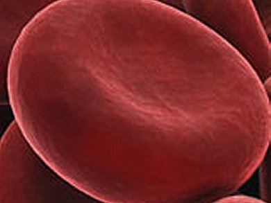
Measuring the composition of a patient’s blood is often the first step in clinical diagnosis, and is most commonly performed by extracting a blood sample for ex vivo analysis using optical flow cytometry and other biochemical assays. This method lacks the ability to provide real-time monitoring of patients in critical conditions.
Lior Golan, Technion-Israel Institute of Technology, Haifa, Israel, and colleagues demonstrate an optical system that allows noninvasive imaging of blood cells in vivo. Their label-free approach for in vivo flow cytometry of blood uses a compact imaging probe that could be adapted for bedside real-time imaging of patients in clinical settings.
With no fluorescence labeling, they show high resolution imaging of red and white blood cells flowing in the oral mucosa (vessel in the lower lip) of a human volunteer. By analyzing the large data sets obtained by the system, valuable blood parameters could be extracted and used for direct, reliable assessment of patient physiology.
This spectrally encoded flow cytometry (SEFC) offers a new set of tools which could be utilized for a wide range of screening and diagnostics applications, and opens new possibilities in the clinical research and practice.
- Noninvasive imaging of flowing blood cells using label-free spectrally encoded flow cytometry,
Lior Golan, Daniella Yeheskely-Hayon, Limor Minai, Eldad J Dann, Dvir Yelin,
Biomed. Optics Express 2012, 3 (6), 1455–1464.
DOI: 10.1364/BOE.3.001455