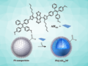
Detecting biomolecules in cells using fluorescence labels attached to an antibody can be tough at low concentrations. To achieve signals that are bright enough, a large amount of fluorophores is needed. However, if they are in close proximity, self-quenching becomes an issue
Krzysztof Matyjaszewski, Bruce A. Armitage, Subha R. Das, and colleagues, Carnegie Mellon University, Pittsburgh, PA, USA, have developed an approach to attach large numbers of fluorophore labels to a single antibody. The researchers used a polymer in the shape of a bottle brush, and attached DNA to the “bristles”. The DNA strands can accomodate the flourescent labels while separating them enough to avoid self-quenching.
The resulting brush-DNA nanotag can be attached to antibodies and detect proteins with a bright signal and high sensitivity. The team points out that their system compares favorably to commercially available antibodies labeled with flourescent dyes. Additionally, the approach could potentially be used to transport DNA intercalating cancer therapeutics.
- Bright Fluorescent Nanotags from Bottlebrush Polymers with DNA-Tipped Bristles,
Munira F. Fouz, Kosuke Mukumoto, Saadyah Averick, Olivia Molinar, Brooke M. McCartney, Krzysztof Matyjaszewski, Bruce A. Armitage, Subha R. Das,
ACS Cent. Sci. 2015.
DOI: 10.1021/acscentsci.5b00259