Chemical Maps
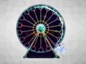
To find our way, we use maps. Cells use “chemical maps” to find the way: they orient themselves by following concentration gradients of attractants or repellants. David H. Gracias and a team at Johns Hopkins University, Baltimore, USA, have now developed a clever new method to produce three-dimensional patterns of chemical concentration gradients in vitro—with previously unattainable versatility and precision in both space and time. As the scientists report, they use tiny containers of different shapes and patterned with different arrangements of slits through which chemical messenger substances can diffuse. They were thus able to induce fluorescing cells to organize themselves into a glowing green spiral.
Concentration gradients not only can guide bacteria, fungi, and amoebae; they are also very important in the early stages of embryogenesis because the development of seed leaves (cotyledon) is controlled through concentration gradients of messenger molecules. Three-dimensional chemical patterns play a role in many physiological and pathological processes, including the growth of blood vessels, regulation of blood pressure and heart rate, and tumor metastasis. Our immune cells also follow concentration gradients to find the spot where they are needed.
3D Concentration Patterns
In order to examine these processes more closely, scientists want to imitate such chemical gradients in vitro. Making a three-dimensional chemical pattern and maintaining it long enough is not so easy. Previous microfluidic methods only allowed for the generation of two-dimensional patterns of limited size. An alternative technique discussed here is the diffusion of chemicals through precisely formed porous containers in stationary media. Variation of the container geometry and pore pattern in the walls makes it possible to realize a wide variety of three-dimensional concentration patterns.
The special trick: Gracias and his co-workers “build” their containers from two-dimensional surfaces held together with tiny hinges. These were designed so that the containers fold up on their own when heated and then stay tightly closed on cooling. In this way, they are able to make containers ranging in size from 100 nm to a few millimeters for potential applications at the sub-cellular to tissue scale. Before being folded, established lithographic methods can be used to perforate each surface with a well-defined arrangement of slits or holes with nano-microscale precision.
With an offset arrangement of slits on four surfaces of a cube shaped container, the researchers were able to release an attractant to generate a concentration gradient in the form of a spiral winding around the container. Fluorescing bacteria followed this pattern and arranged themselves into a glowing spiral.
- Direction of Cellular Self-Organization by the Generation of Three- Dimensional Chemical Patterns
Y. V. Kalinin, J. S. Randhawa, D. H. Gracias,
Angew. Chem. Int. Ed. 2011, 50(11), 2549–2553.
DOI: 10.1002/anie.201007107