Copper, magnesium, and zinc ions are important cofactors in cellular processes. Hua Kuang and colleagues, Jiangnan University, Wuxi, China, have synthesized hybrid chiral nanoprobes which can be used for the imaging and quantitative analysis of these divalent metal ions in living cells.
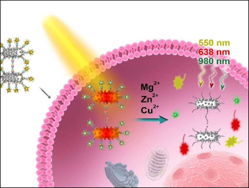
The nanoprobes consist of dimers of spiny Pt-coated Au nanorods attached to upconversion nanoparticles (UNCPs) and two types of fluorescent dyes using complementary DNA strands (pictured top left). The probes show strong circular dichroism. Under left-circular-polarized light illumination at 980 nm, the DNA strands are released due to a rise in temperature caused by a plasmonic photothermal effect (pictured red in the center).
The DNA used contains Zn2+‐, Cu2+‐ and Mg2+‐specific DNAzymes, whose substrates are connected to either one of the fluorescent dyes or the UCNPs. The Zn2+ substrate DNA strand is labeled with the dye cyanine 5 (Cy5), a strand labeled with the dye 5‐carboxytetramethylrhodamine (TAMRA) acts as the Mg2+ substrate DNA strand, and the Cu2+ substrate DNA strand is attached to the surface of the UCNPs.
When the targeted metal ions are present, they recognize their corresponding binding sites and activate the DNAzymes. This causes the DNAzymes to cleave their substrates and free the fluorescent dyes and/or UNCPs, depending on which metals are present. This restores their luminescence signals (pictured right). The luminescence intensities of the released upconversion nanoparticles and the two dyes depend on the concentration of the three metal ions. The probes are highly specific and versatile for the quantitative detection of metal ions in various cells.
- Circular Polarized Light Activated Chiral Satellite Nanoprobes for the Imaging and Analysis of Multiple Metal Ions in Living Cells,
Rui Gao, Liguang Xu, Changlong Hao, Chuanlai Xu, Hua Kuang,
Angew. Chem. Int. Ed. 2019.
https://doi.org/10.1002/anie.201814282