Exosomes are cell-secreted vesicles that carry specific biomarkers from their parent cells. They could allow early diagnosis of cancer. Fluorescence microscopy is commonly used to detect exosomes but the sensitivity of this technique is reduced by autofluorescence in the detection environment.
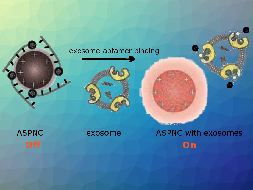
Kanyi Pu and colleagues, Nanyang Technological University, Singapore, have developed a sensor for exosome detection. They used so-called afterglow—an excitation-free luminescence—as the readout signal. The team synthesized a poly(phenylenevinylene)—tetraphenylporphyrin semiconducting polyelectrolyte, which self-assembles into nanoparticles (ASPNs) covered with positive charges. Electrostatic interactions between ASPNs and quencher-tagged aptamers form the sensor complex ASPNC (pictured). The initial afterglow signals for ASPNC are almost quenched. In the presence of exosomes interactions in ASPNCs are disrupted and afterglow signals are turned on (pictured).
Because detection of the afterglow takes place after the excitation, background signals are minimized. This leads to an improved limit of detection that is nearly two orders of magnitude lower than that of fluorescence detection in cell culture media. Detection of different exosomal proteins can be achieved by simply changing the aptamer sequence.
- Near-Infrared Afterglow Semiconducting Nano-Polycomplexes for the Multiplex Differentiation of Cancer Exosomes,
Yan Lyu, Dong Cui, Jiaguo Huang, Wenxuan Fan, Yansong Miao, Kanyi Pu,
Angew. Chem. Int. Ed. 2019, 58, 4983–4987.
https://doi.org/10.1002/anie.201900092