Inorganic nanoparticles with tunable emissions have attracted great interest in biomedical imaging due to their high photochemical stability compared with fluorescent dyes. However, many of the most popular materials contain heavy metals, which has led to concerns about their harmful effects both on the biological samples being studied and the environment in general.
Qin Kuang and colleagues, Xiamen University, China, have developed a route to tune the wavelength of light emitted from heavy-metal-free MgO nanocrystal clusters that relies on the interaction between the inorganic core and organic species at the surface. By modifying the amount of acetic acid added during the synthesis, the emission peaks could be tuned from approximately 400 to 550 nm, with excitation at 365 or 440 nm.
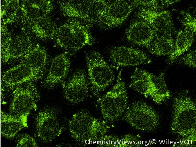
To test the potential application of these nanoparticles in cellular imaging, they were coated with a layer of SiO2 to ensure their stability under cell culture conditions and used for in vitro confocal imaging of HeLa cells. The nanoparticles were found to have high biocompatibility, and a bright signal was observed throughout the cell cytoplasm.
- Organic–Inorganic Interface-Induced Multi-Fluorescence of MgO Nanocrystal Clusters and Their Applications in Cellular Imaging,
Shuifen Xie, Shixiong Bao, Junjie Ouyang, Xi Zhou, Qin Kuang, Zhaoxiong Xie, Lansun Zheng,
Chem. Eur. J. 2014.
DOI: 10.1002/chem.201303927