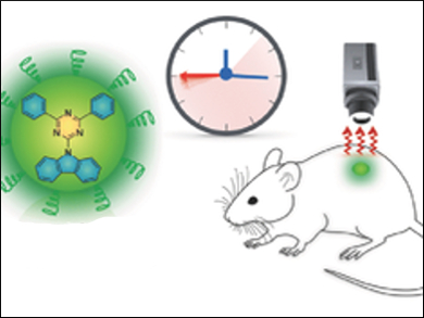
In vivo optical imaging is an important tool in a variety of medical applications, for example, for real-time mapping during surgeries. Most techniques currently need external light excitation during the imaging process. However, this can induce tissue autofluorescence, which affects the image quality. A promising alternative is the so-called persistent luminescence or afterglow, which is luminescence that remains active after removal of the light source. To date, only inorganic nanoparticles made of toxic metals had been reported for in vivo afterglow imaging.
Kanyi Pu, Nanyang Technological University Singapore, and colleagues have developed water-soluble, oxygen-inert organic semiconducting nanoparticles (OSNs). The nanoparticles contain organic phosphorescent semiconducting dyes and are prepared by sonication. The OSNs have long phosphorescence times of more than 10 s when imaging the lymph nodes of live mice without real-time external excitation. Moreover, the phosphorescence of the OSNs persisted over several cycles.
The team demonstrated that the top-down synthesis of the organic nanoparticles from crystalline phosphorescent semiconducting dyes resulted in the formation of aggregates of the dye molecules, in which the dye molecules are aligned parallel to each other. This arrangement stabilizes the triplet excited states, which prolongs the luminescence time of the synthesized OSNs. Luminescence on this time scale can be detected by commercial whole-animal imaging systems.
- Ultralong Phosphorescence of Water-Soluble Organic Nanoparticles for In Vivo Afterglow Imaging,
Xu Zhen, Ye Tao, Zhongfu An, Peng Chen, Chenjie Xu, Runfeng Chen, Wei Huang, Kanyi Pu,
Adv. Mater. 2017.
DOI: 10.1002/adma.201606665