Electron and atom-probe microscopy can categorize atoms in thin sheets of material, and in small areas of thicker samples, but it has proven far more difficult to map the constituents of nanostructures inside large, thick objects. X-rays—the most common imaging tool for hard biological materials such as bones—have a limited focal-spot size, so they cannot focus on nanoscale objects.
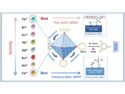
Yukio Takahashi and colleagues at Osaka University, together with researchers at Nagoya University and the RIKEN SPring-8 center in Hyogo, all Japan, have succeeded in producing two-dimensional images of nanostructures encased in thick materials on a large scale. This was possible because they designed a new x-ray diffraction microscopy system, x-ray ptychography, that does not require a lens. Ptychography involves taking images of an object that overlap with one another on a series of coinciding lattice points. This technique was combined with x-rays, and included a system to compensate for the drifting of optics during imaging.
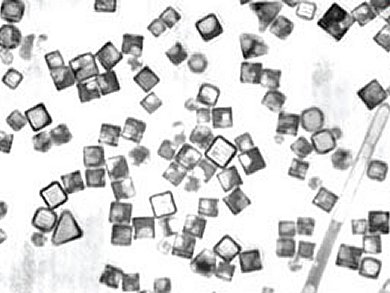
To verify that their system works, the researchers deposited Ag/Au nanoparticles around 200 nm in size on a silicon nitride membrane. They performed quantitative imagings of both the electron density and Au element of Au/Ag nanoparticles at the pixel resolution of better than 10 nm in a field of view larger than 5 × 5 μm2 by directly phasing ptychographic coherent diffraction patterns acquired at two x-ray energies below the Au L3 edge.
This new method to map nanostructures within materials may lead to imaging material and biological systems, e.g., of the internal organization of cells. The shape of a whole cell and the spatial distribution of its organelles could be three-dimensionally visualized at 10 nm resolution.
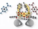
Image: © Yukio Takahashi
- Multiscale element mapping of buried structures by ptychographic x-ray diffraction microscopy using anomalous scattering,
Yukio Takahashi, Akihiro Suzuki, Nobuyuki Zettsu, Yoshiki Kohmura, Kazuto Yamauchi, Tetsuya Ishikawa,
Appl. Phys. Lett. 2011, 99, 131905.
DOI: 10.1063/1.3644396