Do reaction compartments in a cell really need a physical boundary? No, according to Lorenzo Di Michele, Imperial College London, UK, and colleagues, who have transformed a DNA condensate into a microreactor system with reaction and storage compartments. They created an artificial cell containing a “nucleus” for RNA synthesis and a shell for RNA sequestration. No physical boundary separates the compartments, with compartmentalization being purely driven by diffusion and reactivity.
Life Based on Physical Boundaries
Life as we know it is based on cells. Our observations on living cells are regarded as general principles of life. One supposedly fundamental principle is that cells have compartments—such as the nucleus, mitochondria, and the endoplasmic reticulum—which work together in a living cell. These cellular compartments are separated from each other either by a membrane, or they are distinguished by a different phase, as in the case of some cellular bodies like lipid droplets, nucleoli, or granules.
It has long been believed that compartmentalization without a physical boundary, such as a membrane, seems unlikely or even impossible in cells. However, this view is now being challenged by the team. The researchers have succeeded in creating a cell-sized body that makes its own dynamic compartments just by distinguishing between diffusing reagents in terms of their reactivity.
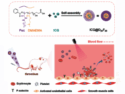
To make this construct, the team designed a DNA condensate forming a homogeneous, but adaptable network. Four DNA strands were locked in a three-dimensional cross to give a four-way junction from which four double-stranded arms pointed outwards. These four arms were capped by cholesterol to obtain an amphiphilic assembly, connected by the cholesterol moieties. One strand, however, had an overhang attached to an oligonucleotide. The sequence of this oligonucleotide was designed as a binding site for incoming oligonucleotide probes. On a larger scale, copies of this DNA condensate assembled as a porous network, forming homogeneous, cell-sized microdroplets in aqueous solution.
Diffusion and Replacement
When the researchers added oligonucleotides to the solution, these oligonucleotides diffused into the network, finding and binding to their matching sites in the condensate. As they moved forward in the droplet, they formed “wavefronts” propagating inwards. More strongly binding oligonucleotides, which were added later, can then replace the original ones, forming another propagating wavefront. The alternating propagation was made visible by the colors of fluorescent dyes attached to the different oligonucleotides (pictured schematically).
However, the team was not satisfied with simply watching wavefronts propagating inward in a droplet. Using special oligonucleotides, they brought the wavefronts to a halt. These stop strands bound most strongly to their matching oligonucleotides and could, therefore, no longer be replaced. With the wavefronts now frozen in their positions, the researchers had established domains, all having the same structure, but different oligonucleotides. These domains could then be used as organelle-like microcompartments, each performing its own chemistry.
“Nucleus” and Shell
To demonstrate the use of the microcompartments, the team created an artificial cell with a “nucleus” and an outer shell domain to produce and store RNA. To prepare the nucleus, the team added oligonucleotides consisting of an RNA template and an RNA polymerase binding site. The shell was made by adding oligonucleotides with a sequence matching that of the RNA produced. Added RNA polymerase then diffused into the nucleus. After binding there, it began producing RNA aptamers, using nucleotides fed to the solution. These aptamers then diffused into the shell, where they bound to their matching oligonucleotides.
This artificial cell is remarkable for two reasons. Firstly, it demonstrates that compartments can evolve through diffusion and reactivity alone, without underlying structural or phase change. This brings into question a basic assumption in synthetic biology, where compartmentalization is mainly associated with vesicle or micelle formation.
It also sheds new light on the action of biological condensates [1] such as lipid droplets or granules, which play a role in living cells. They self-organize and exert function, similar to the DNA condensate produced by the team.
Secondly, the researchers propose exciting uses for DNA condensates in medical biology. They envisage the construction of cell-like agents with a functionality that can evolve over time and space, or domains enriched with molecules to unlock complex reactions, or changing the shape or size of a cell through RNA fluctuations.
Reference
[1] Salman F. Banani, Hyun O. Lee, Anthony A. Hyman, Michael K. Rosen, Biomolecular condensates: organizers of cellular biochemistry, Nat. Rev. Mol. Cell Biol. 2017, 18, 285–298. https://doi.org/10.1038/nrm.2017.7
- Reaction–Diffusion Patterning of DNA-Based Artificial Cells,
Adrian Leathers, Michal Walczak, Ryan A. Brady, Assala Al Samad, Jurij Kotar, Michael J. Booth, Pietro Cicuta, Lorenzo Di Michele,
J. Am. Chem. Soc. 2022, 144, 17468–17476.
https://doi.org/10.1021/jacs.2c06140
![]()