The design of bright/fluorescent probes with biologically benign materials is highly desirable for in vivo clinical purposes.
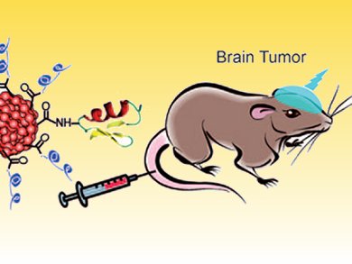
Working with colleagues from China and the USA, Daniel Chiu and co-workers have made an important advance for in vivo imaging based on new organic nanoparticles. They used semiconducting polymer dots—which have exceptional brightness, are nontoxic, and have broad absorption in the visual region with efficient emission in the red region—that were conjugated to a peptide chlorotoxin to specifically target brain tumors in a transgenic mouse model (see figure).
These new bioconjugates are promising candidates as probes for clinical diagnostics.
- Design of Highly Emissive Polymer Dot Bioconjugates for In Vivo Tumor Targeting
C. Wu, S. J. Hansen, Q. Hou, J. Yu, M. Zeigler, Y. Jin, D. R. Burnham, J. D. McNeill, J. M. Olson, D. T. Chiu,
Angew. Chem. Int. Ed. 2011.
DOI: 10.1002/anie.201007461 - C. Wu, S. J. Hansen, Q. Hou, J. Yu, M. Zeigler, Y. Jin, D. R. Burnham, J. D. McNeill, J. M. Olson, D. T. Chiu,
Angew. Chem. 2011.
DOI: 10.1002/ange.201007461