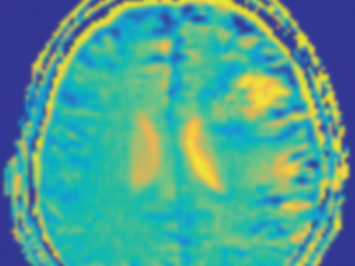
To determine how large tumors are and if they have spread to the surrounding tissue, medical professionals often use diagnostic procedures in which the patient is administered contrast media or exposed to radiation. There is, however, an alternative: Dynamic glucose-enhanced magnetic resonance imaging (DGE-MRI) allows the high-resolution visualization of glucose uptake in the brain without using radioactive isotopes.
Daniel Paech and Patrick Schünke, German Cancer Research Center (DKFZ), Heidelberg, Germany, use the fact that tumor cells have a significantly higher glucose uptake due to the increased cell division rate. Instead of the gadolinium complex used in conventional MRI or the18F-labeled glucose used in positron emission tomography (PET), normal glucose is used as a marker.
The basis for the method is the exchange of hydrogen atoms of the OH groups of the sugar with those of the surrounding free water. This process provides the contrast in the image based on the relative concentration of glucose in different places. The patient lies in a commercially available MRI machine, which, however, has a higher field strength than usual (7 T instead of 1.5–3 T). A glucose solution is administered intravenously. Before, during, and after this injection, the magnetic resonance is measured. The spectra are compared to determine the location and size of the tumor.
In contrast to gadolinium-based contrast agents, which are restricted to the inside of the blood vessel as well as to cell interstices in human tissues, glucose enters the cell interior. As a result, glucose-enhanced imaging can in principle map both the glucose influx via the vessels (perfusion) and intracellular metabolic processes. Studies of healthy subjects and tumor patients indicate that all three compartments, i.e., vessels, intercellular spaces, and the cell interior contribute to the signal.
The results obtained so far show that glucose-enhanced MR imaging is a cost-effective and safe alternative to conventional methods. In addition, it provides more information. The technique could potentially be used to guide the removal of tissue samples from active tumor areas, to plan therapies, and to control the progression of cancers. Further studies in larger populations are needed to further evaluate the potential benefit to clinical routine.
- Glukoseverstärkte Magnetresonanz (in German),
Daniel Paech, Patrick Schünke,
Nachr. Chem. 2018, 66, 496–498.
https://doi.org/10.1002/nadc.20184067174