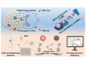
Dermal Tattoo Sensors
The art of tattooing may have found a diagnostic twist. Ali K. Yetisen, Technical University of Munich, Germany, and colleagues have developed permanent dermal sensors that can be applied as artistic tattoos. A colorimetric analytic formulation was injected into the skin instead of tattoo ink. The pigmented skin areas change their color when blood pH or other health indicators change.
A tattoo artist places ink directly in the dermis, a roughly one-millimeter-thick layer of tissue that hosts nerves, blood vessels, and hair follicles. The tattoo needle punctures the epidermis, the uppermost layer of skin, and releases the pigments into the dermis below, where the pigments stain the skin permanently.
Using tattoos for diagnostic rather than cosmetic purposes is a new concept. The team thought the technique could be helpful to place sensor formulations at spots in the body where they can record changes in metabolic substances directly, without any spatial distance or time delay, and perhaps for a very long period of time.
Detection of Blood pH Change and Metabolite Levels
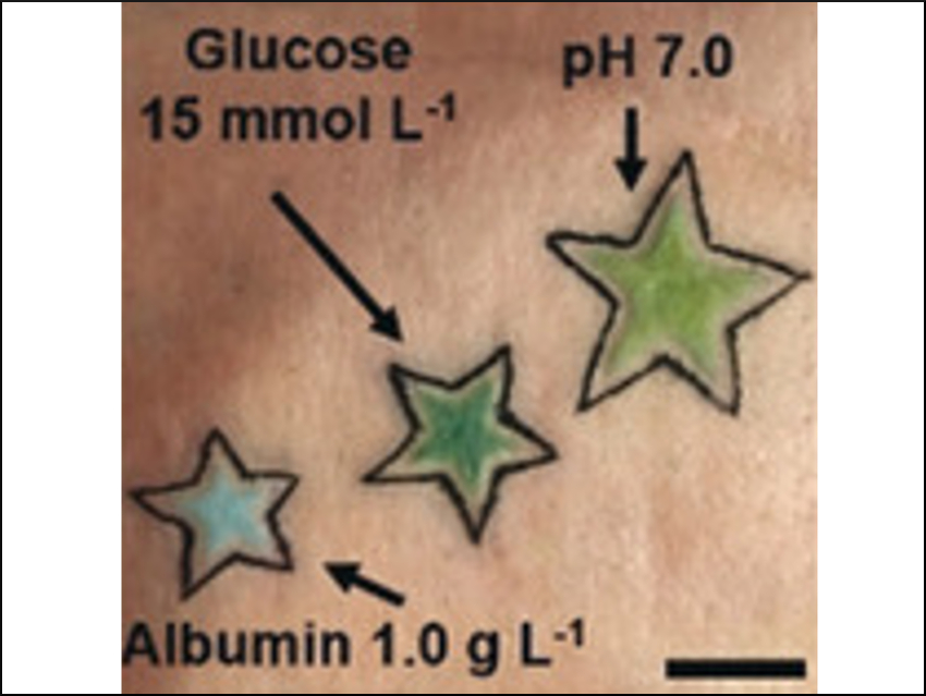
The researchers then identified and adapted three colorimetric chemical sensors that produce a color change in response to biomarkers. The first sensor was a rather simple pH indicator consisting of the dyes methyl red, bromothymol blue, and phenolphthalein. If injected into a model skin patch—a piece of pig skin—the resulting tattoo turned from yellow to blue if the pH was adjusted from five to nine.
The other two sensors probed the levels of glucose and albumin. Albumin is a carrier and transport protein in the blood. High glucose levels in the body may indicate diabetic dysfunction, whereas falling albumin levels can indicate liver or kidney failure. The glucose sensor consisted of the enzymes glucose oxidase and peroxidase, which, depending on the glucose concentration, led to a structural change of an organic pigment, and a yellow to dark green color change. The albumin sensor was based on a yellow dye that, upon association with the albumin protein, turned green.
The scientists then applied several sensor tattoos onto patches of pig skin. When they changed the pH or the glucose or albumin concentrations, the colors of the decorated areas changed accordingly. They quantified these visible effects by evaluating the colors with a simple smartphone camera and an app.
The researchers claim that such sensor tattoos could allow the permanent monitoring of patients using a simple, low-cost technique. With the development of suitable colorimetric sensors, the technique could also extend to recording electrolyte and pathogen concentrations or the level of dehydration of a patient. Further studies will explore whether tattoo artwork can be applied in a diagnostic setting.
- Dermal Tattoo Biosensors for Colorimetric Metabolite Detection,
Ali K. Yetisen, Rosalia Moreddu, Sarah Seifi, Nan Jiang, Katia Vega, Xingchen Dong, Jie Dong, Haider Butt, Martin Jakobi, Martin Elsner, Alexander W. Koch,
Angew. Chem. Int. Ed. 2019.
https://doi.org/10.1002/anie.201904416