Two Imaging Techniques at Once
Magnetic resonance imaging (MRI) visualizes internal body structures, often with the help of contrast agents to enhance sensitivity. Sophie Laurent, University of Mons, Belgium, and Center of Microscopy and Molecular Imaging, Charleroi, Belgium, and colleagues have developed a bimodal contrast agent suited for two imaging techniques at once, namely, MRI and a technique called photoacoustic imaging. The use of only one contrast agent for two imaging techniques improves the sensitivity of both, with only little impact on the patient’s body.
MRI is a widely used technique in medicine and research and is known for its good resolution. Structures down to a hundredth of a millimeter can be resolved. However, sensitivity, the ability to detect something at all, is sometimes an issue. Therefore, contrast agents are often administered to improve the clarity with which the structures can be seen.
The results of an MRI scan can also be improved in combination with complementary imaging methods, which focus on different aspects. However, most imaging tools require the presence of probes and dyes, but applying first a contrast agent, and then a second drug may cause more risks for the patient. This inspired the team to develop so-called bimodal contrast agents—agents that would serve both tools at once.
MRI contrast agents typically contain gadolinium, a paramagnetic element that enhances the signal of the elements nearby. Free gadolinium can be harmful, but it is held tightly within the structure of an organic molecule. Laurent’s idea was to directly join the gadolinium agent with the probe used for the second imaging technique.
MRI and Photoacoustic Imaging
The team chose photoacoustic imaging (PAI), a highly sensitive and rather new imaging method that measures the heat response in a tissue to laser pulses. The method is non-invasive like MRI, but a special organic dye must be present that absorbs laser light applied from the outside. This technique would clearly enhance MRI sensitivity, the team thought. Dysfunctions in the skin and below would be detected with unprecedented clarity.
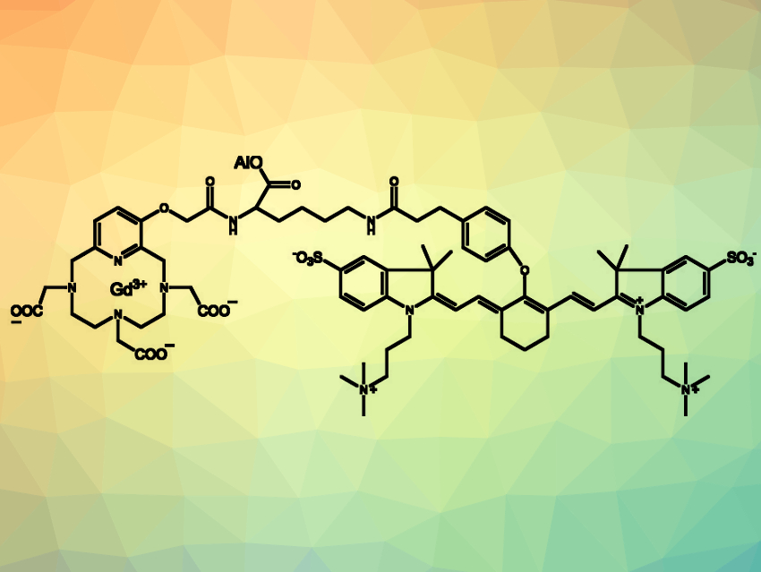
To join the gadolinium agent with the organic dye, the scientists chose the natural amino acid lysine as a linker. Lysine is special among the amino acids. It is a rather long molecule that can bind to two other molecules at both ends. The scientists successfully linked an MRI agent called Gd-PCTA with a PAI probe called ZW800-1. Apart from the two connections, lysine has a third possible connectivity, which could be of use in the future. The scientists imagine adding an additional biovector, for example, a peptide that recognizes a biological disorder—this would make the now bimodal probe trimodal.
The bimodal probe enhanced the MRI contrast as strongly as a commercial MRI agent and gave the same photoacoustic signal as the original PAI probe. This means the probe is a two-in-one agent, which facilitates the combination of MRI and other medical imaging techniques. The next step would be to test it in real organisms.
- Bimodal Probe for Magnetic Resonance Imaging and Photoacoustic Imaging Based on a PCTA-Derived Gadolinium(III) Complex and ZW800-1,
Marie Devreux, Céline Henoumont, Fabienne Dioury, Dimitri Stanicki, Sébastien Boutry, Lionel Larbanoix, Clotilde Ferroud, Robert N. Muller, Sophie Laurent,
Eur. J. Inorg. Chem. 2019, 2019, 3354–3365.
https://doi.org/10.1002/ejic.201900387