Noninvasive imaging of β cells could help understanding islet formation and the pathophysiology of diabetes. Progressive loss of functional β cell mass is the underlying cause of autoimmune type 1 diabetes mellitus. It is also responsible for the secondary failure of clinical drugs in type 2 diabetes.
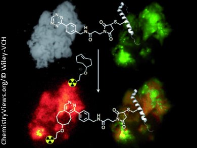
Ralph Weissleder and co-workers, Harvard Medical School, Boston, USA, used a cysteine at position 12 (Cys12) to label an exendin-4 analogue, which showed sub-nanomolar EC50 binding concentrations and specificity to glucagon-like peptide 1 (GLP-1) receptors on β cells, with 18F. Thereby they make the exendin-4 analogue suitable for positron emission tomography (PET) imaging. Cys12 was modified with a tetrazine via a maleimide-NHS ester, and 18F was then introduced based on the transcyclooctene/tetrazine cycloaddition.
The resulting PET imaging agent accumulated in pancreatic β cells and in different murine xenografts of insulinomas in mice. An uptake could be inhibited by unlabeled exendin-4. Pharmacokinetic modeling and allometric scaling predicted a clearance half-life of 18 min in human, and, with the decay of 18F being 109.8 min, this gives the 18F-modified exendin-4 close to ideal clearance properties for human imaging.
 Efficient 18F-Labeling of Synthetic Exendin-4 Analogues for Imaging Beta Cells,
Edmund J. Keliher, Thomas Reiner, Greg M. Thurber, Rabi Upadhyay, Ralph Weissleder,
ChemistryOpen 2012, 1(4), 177–183.
DOI: 10.1002/open.201200014
ChemistryOpen – the first society-owned, open-access, chemistry journal – is a journal of ChemPubSoc Europe published by Wiley-VCH.