Direct potentiometric determination of the sialic acid concentration on cell surfaces—a new technique for tumor diagnosis?
Quick and Easy Differentiation
For proper cancer treatment, it is critically important to know the extent of the disease. Any operation should completely remove all affected tissue. In order to catch stray cells that may have already moved on into healthy tissue it is necessary to take out healthy adjacent tissue and sometimes other affected organs and lymph nodes. Japanese researchers working with Yuji Miyahara have now developed a technique that allows for the quick and easy differentiation between diseased and healthy tissue. As the scientists report in the journal Angewandte Chemie, the method is based on the direct potentiometric measurement of a tumor marker on cell surfaces.
The cells in our bodies have chains made of special sugar components on their surfaces. Sialic acid is one of these sugar building blocks and is often found at the ends of the sugar chains. These sugar chains can serve as a signal for the detection of certain pathological processes. For example, certain types of cancer involve an overproduction of sialic acid in the tumor cells, which causes these molecules to build up in the cell membrane. This increase in membrane-bound sialic acid can be detected in blood serum and is occasionally used as a test for the early detection of cancer.
Specific Binding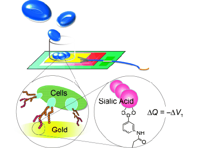
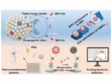
The research team from the University of Tokyo and National Institute for Materials Science (Japan) has now developed an interesting new method for the detection of elevated sialic acid levels that can quickly, easily, and directly determine whether a tissue sample contains malignant mutated cells, and how far the metastasis of a tumor has progressed. For their potentiometric process, the researchers use the fact that sialic acid specifically binds to a compound named phenylboronic acid (PBA). Related sugar molecules do not bind to PBA. The scientists coated gold electrodes with a layer of PBA. If the coated electrodes come into contact with a sample that contains cells with sialic acid, the cells bind to the PBA through their sialic acid molecules. Cells that contain many sialic acid molecules bind more strongly to the electrode. This changes the electrical properties of the electrode. Changes in surface potential are then monitored and used to quantify the sialic acid concentration in the sample.
To carry out the test, a sample of the suspicious tissue must simply be extracted and suspended. The number of cells contained in a defined volume of the suspension is then determined, and the suspension is poured over the electrode. No other sample preparation is required. Carried out in parallel to established histological tests, the potentiometric examination could rapidly deliver supplementary information about the malignancy of tumors and the degree of metastasis.
- Assessment of Tumor Metastasis by the Direct Determination of Cell-Membrane Sialic Acid Expression
A. Matsumoto, H. Cabral, N. Sato, K. Kataoka, Y. Miyahara,
Angew. Chem. Int. Ed. 2010, 49 (32), 5494 – 5497.
DOI: 10.1002/anie.201001220 - A. Matsumoto, H. Cabral, N. Sato, K. Kataoka, Y. Miyahara,
Angew. Chem. 2010, 122 (32), 5626 – 5629.
DOI: 10.1002/ange.201001220