Specific Visualization of Cartilage Tissue
The mainstream techniques for visualization of cartilage tissue in the body are magnetic resonance imaging and computer tomography, but both techniques do not provide optimum quality images. A new method projected by American and South Korean scientists is based on contrast agents that can be detected in the near-infrared (NIR) wavelength range. Injection of the fluorophores allows high-quality visualization of cartilage tissue in real time, as the authors report in the journal Angewandte Chemie.
Advantages of Near-Infrared Fluorophores
Using NIR fluorophores for tissue imaging has several advantages. Hak Soo Choi from Harvard Medical School, Boston, USA, and Hoon Hyun from Chonnam National University, South Korea, explain: “The most important advantage of using NIR fluorophores for cartilage imaging is the integrated method of performing ‘structure-inherent targeting’ (i.e. molecular targeting and fluorescent imaging) using a single small molecule. The high affinity of cationic NIR fluorophores for cartilage provides high-quality tissue visualization, ultralow background signal, and low injected dose”.
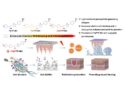
Therefore, the Choi group in collaboration with Maged Henary at Georgia State University, Atlanta, USA, screened an NIR fluorophore library for their cartilage specificity. The best fluorophores were based on a polymethine core with quaternary ammonium cations. They were re-synthesized and chemically altered to fine-tune their physicochemical properties. Two highly promising molecules were then successfully tested in mouse as well as Yorkshire pig models for their tissue-imaging properties.
Detecting Cartilage and Bone Simultaneously
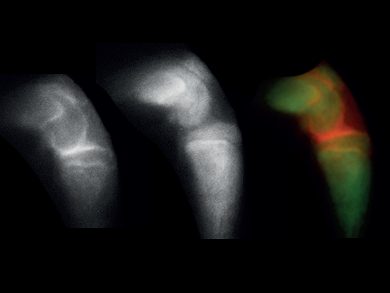
Although it is not very clear yet what the actual binding target of the contrast agents is, the scientists believe that the highly negatively charged components of the cartilage tissue are the targets that are recognized and specifically bound by the positively charged fluorophores. In addition, the new NIR fluorophores are very stable and not degraded in the body, and the strong cartilage signal was obtained 1-4 hours after the injection of a single dose. Furthermore, the NIR fluorophores also offer the possibility of detecting cartilage and bone tissue simultaneously in real time by a dual-NIR-channel method based on the recently developed bone-specific NIR fluorophores in combination with the cartilage-specific NIR fluorophores.
NIR fluorophores not only have advantages over the established mainstream imaging techniques, but also surpass other methods based on other contrast agents, as Choi explains: “NIR fluorophores provide high spatial resolution in real time to distinguish the boundary and thickness of cartilage tissues from the adjacent bones, where other contrast agents with nanoparticle formulations have limited penetration and delineation”. Thus, the new NIR cartilage-specific fluorophores may be highly beneficial in many applications including investigation of cartilage degeneration, new drug design, and image-guided surgery.
- Cartilage-Specific Near-Infrared Fluorophores for Biomedical Imaging,
Hoon Hyun, Eric A. Owens, Hideyuki Wada, Andrew Levitz, GwangLi Park, Min Ho Park, John V. Frangioni, Maged Henary, Hak Soo Choi,
Angew. Chem. Int. Ed. 2015.
DOI: 10.1002/anie.201502287
Also of Interest
- Phosphonated Near-Infrared Fluorophores for Biomedical Imaging of Bone,
Hoon Hyun, Hideyuki Wada, Kai Bao, Julien Gravier, Yogesh Yadav, Matt Laramie, Maged Henary, John V. Frangioni, Hak Soo Choi,
Angew. Chem. Int. Ed. 2014, 53, 10668–10672.
DOI: 10.1002/anie.201404930