Traditionally, the detection of different cell types, such as cancerous and non-cancerous, is achieved through fluorescence-based techniques. While these techniques are well characterized, the ability to run multiple assays is limited by broad signals. Surface-enhanced Raman spectroscopy (SERS) has recently emerged as an alternative to fluorescence-based techniques for its multiplexing capabilities. However, its use is often hindered by signal variation and strong fluorescence background.
Gilbert Walker, University of Toronto, Canada, and colleagues have developed a strategy to generate an extremely strong Raman signal with reduced fluorescence. Using a cationic linker layer, the team attached a J-aggregate forming dye onto monodisperse gold nanoparticles. The resultant system was encapsulated in either phospholipid (control) or silica.
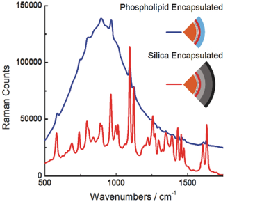
The researchers found that once encapsulated in silica, the fluorescence background was attenuated by five times and the Raman scattering was enhanced by seven times compared to the control (see picture). The exhibited signal was 24 times greater than comparable commercial standards. The group attributes this result to proximal gold quenching by the gold as well as better packing of the dye as a result of the rigid silica shell. The researchers suggest that the combination of reduced fluorescence background, enhanced SERS intensity, and temporal stability makes these particles highly attractive for high-throughput applications.
- Bright Surface-Enhanced Raman Scattering with Fluorescence Quenching from Silica Encapsulated J-Aggregate Coated Gold Nanoparticles,
Christopher M. Walters, Caroline Pao, Brandon P. Gagnon, Colin R. Zamecnik, Gilbert C. Walker,
Adv. Mater. 2017.
https://doi.org/10.1002/adma.201705381