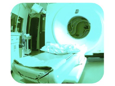
Glioblastoma, the most aggressive form of brain cancer, requires surgical resection. To maximize the accuracy of this procedure, magnetic resonance (MR) and fluorescent-based imagining methods are widely used. Upconversion nanoparticles (UCNPs), namely nanoparticles converting near infrared radiation into visible light, are promising bimodal imaging contrast agents as they provide both MR and fluorescent images of cancerous tissues. Nevertheless, they only poorly penetrate the brain–blood barrier (BBB), a barrier separating the blood from the tissues of the central nervous system. Thus, the use of these nanoparticles for operating on glioblastomas remains challenging.
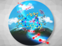
To overcome this issue, Jianlin Shi, Chinese Academy of Science, Shanghai, China, and co-workers synthesized gadolinium-doped UCNPs, coated them with poly(ethylene glicol) and covalently coupled them with Angiopep-2. As this molecule binds to the low-density lipoprotein-receptor-related protein, a protein abundant in the BBB and gliobastoma cells, the new nanoprobes (ANG/PEG-UCNPS) could efficiently cross the BBB and reach tumor cells. They provided accurate MR and fluorescent imagining of glioblastomas without showing toxic effects. Thus, ANG/PEG-CUNPs hold great potential for dual gliobastoma imaging.
- Dual-Targeting Upconversion Nanoprobes across the Blood–Brain Barrier for Magnetic Resonance/Fluorescence Imaging of Intracranial Glioblastoma,
Dalong Ni, Jiawen Zhang, Wenbo Bu, Huaiyong Xing, Fang Han, Qingfeng Xiao, Zhenwei Yao, Feng Chen, Qianjun He, Jianan Liu, Shengjian Zhang, Wenpei Fan, Liangping Zhou, Weijun Peng, Jianlin Shi,
ACS Nano 2014, 8, 1231–1242.
DOI: 10.1021/nn406197c