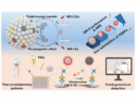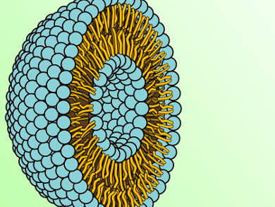
Cell membranes are dynamic structures which release extracellular vesicles (EVs) upon activation or apoptosis. These tiny sub-μm size particles are found in blood and other unprocessed body fluids and take part in various biological properties and functions. Elevated levels of EVs have been reported present in clinical samples from patients with cardiovascular diseases, cancer, sepsis, and auto-immune diseases, making EVs suitable biomarkers for their diagnosis and possibly treatment.
Conventional flow cytometry (FCM) based on light scattering triggering has been used to study EVs for the last twenty years. However, the current technical limitations of this method prevent it from accurately detecting and quantitating EVs of particularly small sizes. Alain Brisson and coworkers, University of Bordeaux-IPB, Pessac, France, have compared the conventional FCM approach to an alternative method that uses fluorescence triggering. In this study, the alternative FCM detected 50 times more Annexin-A5 binding EVs (Anx5+ EVs) in plasma than the conventional FCM. The researchers enumerated and phenotyped EVs from platelet free plasma, focusing on CD41+ and CD235a+ EVs, as well as their Anx5+ and Anx5– sub-populations, which produced similar results.
The researchers point out that this application is simple, sensitive, and reliable, and will help uncover the mechanisms of formation and the role of EVs in different pathologies, subsequently making them reliable disease biomarkers.
- Fluorescence triggering: A general strategy for enumerating and phenotyping extracellular vesicles by flow cytometry,
Nicolas Arraud, Céline Gounou, Delphine Turpin, Alain R. Brisson,
Cytometry A 2015.
DOI: 10.1002/cyto.a.22669