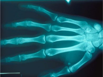
X-rays have been used in biomedical imaging for over a century and nowadays the CT (computed tomography) scan is a common tool for 3D imaging in hospitals.
Pierre Thibault and co-workers, Technische Universität München, Germany, report the generation of 3D electron density maps with high contrast by using a ptychographic coherent imaging approach. The technique uses a monochromatic, pencil X-ray beam to scan the sample, rather than a beam focused by a lens. The sample diffracts the beam, and the intensity of the transmitted beam and its full diffraction pattern are collected. The 3D image is constructed from these 2D projections by using CT-based algorithms.
With this technique, the team has visualized the density differences in a mouse’s femur to display the canalicular network of the tissue within the surrounding bone matrix.
- Ptychographic X-ray computed tomography at the nanoscale
M. Dierolf, A. Menzel, P. Thibault, P. Schneider, C. M. Kewish, R. Wepf, O. Bunk, F. Pfeiffer,
Nature 2010, 467, 436–439.
DOI: 10.1038/nature09419


