Gold nanoparticles are well suited to biomedical applications due to their facile synthesis and relative inertness. Labeling the particles with radionuclides allows them to be used as a probe for single photon emission computed tomography (SPECT) or positron emission tomography (PET), which are used to detect and diagnose cancer. Typically, the radionuclides are bound to the nanoparticles using chelating ligands. However, the stability of these probes is not sufficient for accurate image-guided cancer treatment.
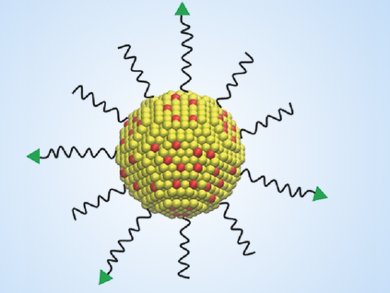
Younan Xia, Georgia Institute of Technology and Emory University, Atlanta, USA, Yongjian Liu, Washington University School of Medicine, St. Louis, MO, USA, and colleagues have incorporated the radionuclide 199Au directly into gold nanoparticles, thereby improving the radiolabel’s stability. To achieve this, the researchers added 199Au3+ ions to the reaction solution during the growth process of the nanoparticles.
The resulting doped nanoparticles were tested in a mouse model for breast cancer and allowed high-quality SPECT imaging. Additionally, the researchers were able to funtionalize the nanoparticles’ surface with D-Ala1-peptide T-amide (DAPTA), which binds specifically to the CCR5 receptor, which is a useful marker for aggressive and metastatic cancer cells. According to the team, this imaging approach could have potential for the sensitive detection of both primary tumors and metastases and help to improve treatment.
- Gold Nanoparticles Doped with 199Au Atoms and Their Use for Targeted Cancer Imaging by SPECT,
Yongfeng Zhao, Bo Pang, Hannah Luehmann, Lisa Detering, Xuan Yang, Deborah Sultan, Scott Harpstrite, Vijay Sharma, Cathy S. Cutler, Younan Xia, Yongjian Liu,
Adv. Healthcare Mater. 2016.
DOI: 10.1002/adhm.201500992