
Carbon dots (CDs) are carbon nanoparticles which have interesting and useful fluorescence properties. They can be applied, e.g., in optoelectronics and bioimaging, particularly if they show long-wavelength emissions (orange to red light). However, such CDs often require complicated syntheses and have broad emission peaks.
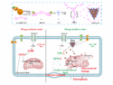
Ye Sun and Miao Yu, Harbin Institute of Technology, China, and colleagues have developed a simple, room-temperature synthesis of nitrogen-doped carbon dots. The team used a one-pot method, combining sodium naphthalene and acetonitrile in ethylene glycol dimethyl ether. The synthesized CDs are hydrophobic, but can easily be modified to be water-soluble by using a polysorbate surfactant.
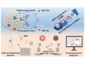
The nitrogen-doped carbon dots were characterized using transmission electron microscopy (TEM) and atomic force microscopy (AFM). They are monodisperse with an average diameter of 2.6 nm. The CDs show an emission at about 588 nm with a narrow peak and a photoluminescence quantum yield of 15 %. The researchers tested their suitability for bioimaging in vitro and in vivo and found them to be an effective and biocompatible imaging agent.
- Nitrogen-doped carbon dots with excitation-independent long-wavelength emission produced by room-temperature reaction,
Chenhui Yang, Shoujun Zhu, Zhenglin Li, Zhuo Li, Chong Chen, Lei Sun, Wei Tang, Rui Liu, Ye Sun, Miao Yu,
Chem. Commun. 2016.
DOI: 10.1039/c6cc06673a

A simple, room-temperature synthesis of nitrogen-doped carbon dots