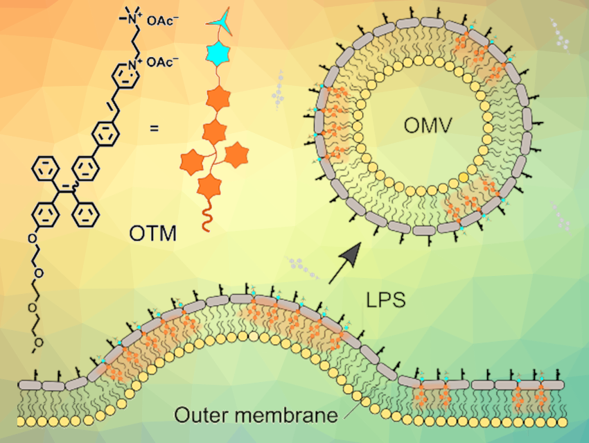
Under stress conditions, Gram-negative bacteria secrete outer membrane vesicles (OMVs) with diameters of 20–250 nm. These small spherical structures carry a series of bacterial components like lipopolysaccharides (LPS) and DNA, and are actively involved in many processes such as quorum sensing, stress responses, etc. OMVs from symbiotic bacteria can affect their hosts—e.g., the immune system. OMVs could also be useful as carriers in therapies against cancer and other diseases. Methods for the detection and quantification of OMVs are, thus, useful.
Engui Zhao, Harbin Institute of Technology, Shenzhen, China, Sijie Chen, Karolinska Institutet, Hong Kong, China, and colleagues have developed an AIE-active fluorescence “turn-on” probe called OEO-TPE-MEM (or OTM) for imaging and quantitative analysis of OMVs. OTM has a very weak affinity towards Gram-negative bacteria in buffer saline solutions, and thus, emits only faintly. The presence of an OMV inducer, such as ampicillin, leads to the remodeling of the bacterial membrane and OMV secretion, which efficiently turns on the fluorescence of OTM. By tracking the fluorescence intensity of OTM, information on the quantity of OMVs can be obtained.
Based on this principle, the researchers established a fluorescence-based technique for the high-throughput detection and identification of OMVs. Compared with conventional biochemical methods such as differential centrifugation, ultrafiltration, dynamic light scattering, or transmission electron microscopy, the reported method has advantages such as high sensitivity and accuracy, as well as easy and fast operation. This high-throughput screening method could provide a useful tool, e.g., for the evaluation of the side effects of antimicrobial materials.
- An AIE‐active probe for efficient detection and high‐throughput identification of outer membrane vesicles,
Fei Wang, Po‐Yu Ho, Chuen Kam, Quanzhi Yang, Jinzhao Liu, Weiping Wang, Engui Zhao, Sijie Chen,
Aggregate 2023.
https://doi.org/10.1002/agt2.312
![]()