Human tear fluids contain many proteins, metabolites, and other molecules whose concentrations change significantly with certain diseases. Yun Feng, Peking University, Third Hospital, Beijing, China, Zhou Yang, University of Science and Technology Beijing, China, Tie Wang, Tianjin University of Technology, China, and Institute of Chemistry, Chinese Academy of Sciences (CAS), Beijing, and colleagues have developed a handy test kit for tears that can identify patients with jaundice. Their success is based on a hybrid sensor that simultaneously removes impurities from the sample. This approach could provide new methods for early detection and diagnosis based on complex bodily fluids.
Tear Fluid Diagnosis
One particular advantage of tear fluid diagnosis is that samples can be collected in a comfortable and non-invasive manner. Surface-enhanced Raman spectroscopy (SERS) is a suitable candidate for the analysis of the biomolecules obtained. The Raman effect is a phenomenon in which light striking materials causes characteristic vibrations and rotations of molecular fragments. The resulting shift in the frequency of the scattered light gives a molecular “fingerprint”.
If the analyte molecules are in contact with a metal surface (hotspots), the Raman signals are amplified enough to reach the ultra-sensitivity required for tear diagnostics. Labeling the analytes is unnecessary. Compact Raman hand devices would be available for direct diagnosis in the field. The problem is in finding suitable SERS sensors. Current sensors are quickly deactivated by the deposition of tear components. Is there a way to make this work without complex sample preparation?
Sensor with Separation Capability
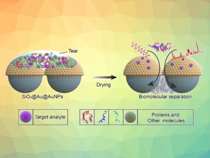
In fact, there is. The team demonstrated this with their novel matchbox-sized diagnostic test kit. At its heart lies a hybrid film. A layer of symmetrically arranged silicon dioxide nanospheres is coated with a thin layer of gold, onto which is deposited a layer of gold nanoparticles that act as SERS hotspots. The target molecules bind to the gold and are held fast to the surface, while smaller tear components slip through the gaps between the silicon dioxide nanoparticles onto an absorbent layer below (pictured).
The pore diameters can be adjusted by changing the size of the nanospheres to selectively separate out the disruptive primary components of tears (albumin, lysozyme, IgG, and peroxidase). The film, called SiO2@Au@AuNPs, is sandwiched between two glass supports and enclosed in a housing. A tip protrudes from the device to collect tear fluid from the corner of the eye. The SERS analysis occurs through a window on top of the device.
The team was able to successfully identify patients with jaundice—a metabolic disorder associated with liver and gall bladder diseases. The bile pigment bilirubin is not properly excreted from the body, becomes concentrated, and can be found in tear fluid. Bilirubin binds strongly to the sensor’s gold and can be detected with great sensitivity by its SERS signal.
- A Separation‐Sensing Platform Performing Accurate Diagnosis of Jaundice in Complex Biological Tear Fluids,
Weidong Zhao, Jinming Li, Zhenjie Xue, Xuezhi Qiao, Ailin Li, Xiangyu Chen, Yun Feng, Zhou Yang, Tie Wang,
Angew. Chem. Int. Ed. 2022.
https://doi.org/10.1002/anie.202205628
![]()