The quick assessment of trace blood components in untreated blood samples is possible with fluorescence tests but, in practice, the blood’s strong autofluorescence interferes with the analysis. Hongwen Liu, Ronghua Yang, Hunan Normal University, Changsha, China, and colleagues have introduced a novel fluorescence probe that quenches this autofluorescence and precisely quantifies traces of hydrogen sulfide, which is an important signal molecule.
Important Trace Analytes
Some blood components are present in extremely low concentrations but are anything but unimportant. For example, the toxic gas hydrogen sulfide (H2S), which smells of rotten eggs, is an important messenger molecule in the body. It is, among other things, involved in regulating circulation. Patients with cardiovascular diseases usually have a reduced H2S concentration in their blood, whereas patients with colon cancer often have elevated levels. The quantification of this and other trace components is, thus, helpful in forming a diagnosis and for the investigation of physiological and pathological relationships.
Fluorescence tests are especially powerful in the analysis of biomolecules. They are inexpensive, uncomplicated, highly sensitive, and can be used for real-time measurements. The problem is that trace components cannot be detected in whole blood samples because blood itself strongly fluoresces, overwhelming weaker signals. Whole blood is, therefore, centrifuged and only the plasma is analyzed. However, this reduces the concentrations of unstable components and gas molecules, yielding inaccurate results.
Autofluorescence Suppression Strategy
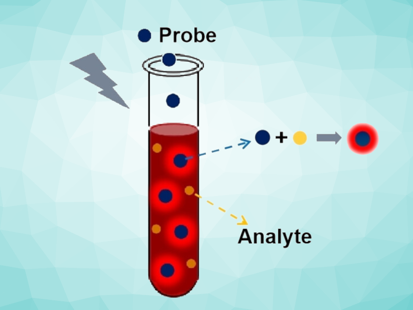
The team developed a novel technique for fluorescence tests that can successfully quantify H2S in whole blood. The trick is that the fluorescence probe itself almost completely quenches the interfering autofluorescence of the blood. The fluorescence dye very strongly absorbs the light that is given off by the autofluorescence of blood components.
The fluorescence dye the researchers used is based on a borodipyrromethene (BODIPY) unit. This quencher has an absorption wavelength in the range of 600–700 nm and high quenching efficiency. It was modified by the addition of two molecular fragments that “recognize” H2S, i.e., two 7-nitrobenzo[c][1,2,5]oxadiazole ether groups. The presence of H2S removes these groups and activates the probe, which begins to fluoresce. Because the autofluorescence of the blood is quenched at the same time, the background fluorescence remains very low and does not interfere.
Accurate Quantification of H2S
Fluorescence tests using the new probe on whole blood samples from patients with cardiovascular diseases verified their reduced H2S levels. Tests on mice with colon cancer showed, also as expected, elevated blood H2S concentrations. According to the researchers, the developed probe is the first fluorescent molecular probe that has achieved the analysis of endogenous H2S in whole blood samples.
In addition, red blood cells from mice were treated with allicin. Allicin is the fragrant compound in garlic that is also responsible for its positive medicinal effects, such as reducing blood pressure. The team was able to use their new probe to demonstrate that allicin triggers the formation of H2S in red blood cells. The researchers hope to use their strategy to develop additional probes for other trace analytes in whole blood.
- A Quencher‐Based Blood‐Autofluorescence‐Suppression Strategy Enables the Quantification of Trace Analytes in Whole Blood,
Zhiyang Yuwen, Qin Zeng, Qiaozhen Ye, Yixing Zhao, Jingxuan Zhu, Kang Chen, Hongwen Liu, Ronghua Yang,
Angew. Chem. Int. Ed. 2023.
https://doi.org/10.1002/anie.202302957
![]()