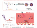
Virus pseudotyping is an analytical method that allows scientists to study virus–cell interactions without the risk of infection associated with the exposure to “real” viruses. It is useful for developing new drugs, finding new ways to treat viral infections such as COVID-19, and testing treatments that have been developed.
Markus Sauer, Gerti Beliu, University of Würzburg, Germany, and colleagues have combined virus pseudotyping with genetic code expansion and click chemistry to create a precise and reliable way to visualize and track infections with a pseudovirus that contains SARS-CoV-2 spike proteins. The work greatly improves on current visualization techniques and provides a more realistic view of virus–cell interactions [1].
Virus Pseudotyping
Virus pseudotyping allows scientists to study highly infectious, often dangerous, viruses without needing to use specialist biosafety facilities. Viral surface proteins, for example, the well-known spike protein of SARS-CoV-2, are transferred to a viral vector without the genetic information needed to replicate the entire virus during infection. This produces a pseudovirus that can be used to study virus–host interactions and also allows scientists to screen for neutralizing antibodies or to test other therapeutics.
In current pseudotyping techniques, a pseudoviral version of vesicular stomatitis virus (VSV) attached to green fluorescent protein (GFP) is used to track infection. After the pseudovirus has infected the cell, it hijacks the cell’s protein-producing machinery to translate its genetic code into viral proteins. Because the genetic code now includes instructions to make GFP, visualizing the characteristic green fluorescence of this protein indicates successful infection, which can be monitored simply by counting fluorescing cells.
Immunolabeling and Its Challenges
Visualization by immunolabeling is another common method for tracking virus–cell interactions. Immunolabeling is based on the use of fluorophore-functionalized antibodies that bind to specific antigens. However, this can sometimes interfere with the natural process of infection, not giving an accurate representation of the impact of the virus in living cells.
Tags or labels added to the virus to track infection can even alter the specificity of the virus, again resulting in pseudoviruses that do not really mimic the actual infectivity of the virus in question.
Combining Pseudotyping, Genetic Code Expansion, and Click Chemistry
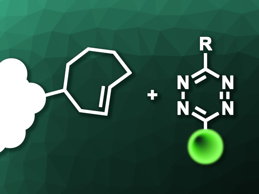
Genetic code expansion (GCE) is the addition of unnatural amino acids, i.e., ones that do not occur in nature, into target proteins at specific sites of interest. The team used genetic code expansion to access three specific sites in the SARS-CoV-2 spike protein and insert trans-cyclooctene (TCO)-functionalized lysine as an amino acid that can be used in click reactions.
Click chemistry uses groups that react highly specifically and quickly with one another in one-pot processes. In the case of the team’s pseudovirus, the trans-cyclooctene group can react with fluorescent compounds that feature tetrazine groups (pictured) in a very fast cycloaddition reaction.
The fluorescent labels can then be used to visualize the pseudoviruses. Importantly, the click reactions are bioorthogonal, meaning they do not interfere with biological processes, including the viral infection of cells. The team tested the infectivity of the pseudovirus with the expanded genetic code, which they named “clickVSV”, alongside uninfected cells and cells infected with an “unclickable” wild-type version of SARS-CoV-2 with conventional GFP labeling. This test showed that clickVSV was as good as the conventionally labeled pseudovirus at infecting cells. They also tested whether virus infectivity was affected by adding the click dyes, finding no noteworthy differences.
The researchers also wanted to test whether infection with clickVSVs would be inhibited in the same way as wild-type viruses, and found no change, meaning their system is also suitable for studying agents that could inhibit or neutralize viruses.
Efficiency of Click Labeling Versus Immunolabeling
The team used two-color dSTORM fluorescence microscopy, counting the number of spike proteins on pseudovirus particles detected by click labeling, which fluoresced magenta, or immunolabeling, which fluoresced cyan. They found, somewhat shockingly, that click-labeling enabled the detection of roughly twice the number of SARS-CoV-2 spike proteins than immunolabeling. The researchers highlight that this not only shows the increased precision of their technique, but also suggests that conventional immunolabeling has been regularly underestimating the amount of viral surface proteins present.
The researchers also visualized the infection of HEK cells with clickVSVs using live-cell lattice-light-sheet (LLS) microscopy in real-time, achieving more than one hour of live 3D imaging of infection. LLS uses a sheet of light to excite fluorescence within the dyes in the clickVSVs one layer at a time, building up a 3D picture in a process akin to the slices taken in an MRI. They also used this type of microscopy to track the movement of individual clickVSVs over the surface of a cell membrane. Compared to particles tracked using immunolabeling, the team again observed that click labeling led to more particle tracks being identified.
Potential Future Uses
The team sees potential uses for clickVSVs in neutralization assays (testing viruses for neutralizing antibodies), investigating viral tropism (the range of cells or cell types that a given virus will infect), receptor binding, and virus-host interactions. The new system, combining genetic code expansion, VSV pseudotyping, and click chemistry labeling is simple, cost-effective, and produces more precise results than conventional methods. The team also highlights that this system can be easily modified to study other virus surface proteins or mutations.
Reference
[1] Re-Engineered Pseudoviruses for Precise and Robust 3D Mapping of Viral Infection,
Marvin Jungblut, Simone Backes, Marcel Streit, Georg Gasteiger, Sören Doose, Markus Sauer, Gerti Beliu,
ACS Nano 2023, 17, 21822–21828.
https://doi.org/10.1021/acsnano.3c07767
Update (December 13, 2023):
A part of the article had originally not been uploaded; this has been corrected.
![]()