|
Entry 1 |
Entry 2 |
|||
 |
 |
|||
|
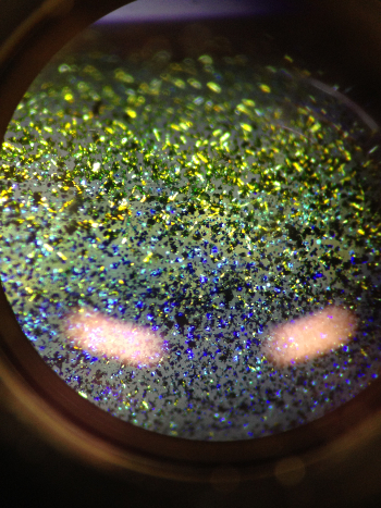
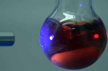
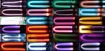
“Visual Signatures” Photo by Spiros Kitsinelis The visual signatures of different gases and vapors in low-pressure discharges |
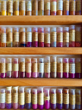
“50 Shades of Cobalt” Photo by Frank T. Edelmann Collection of cobalt(III) complexes
|
|||
|
|
||||
|
Entry 3 |
Entry 4 |
|||
 |
 |
|||
|
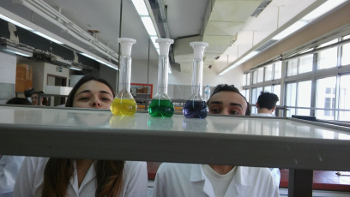
“Curiosity” Photo by Anastasios Papavasileiou Maybe curiosity killed the cat—the scientist still lives with it! |
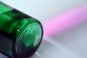
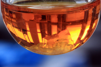
“Complementary Colors” Photo by Julia Bader Reflection of a window frame in a flask with crystals |
|||
|
|
||||
|
Entry 5 |
Entry 6 |
|||
|
|
 |
|||
|
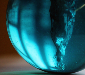
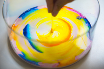
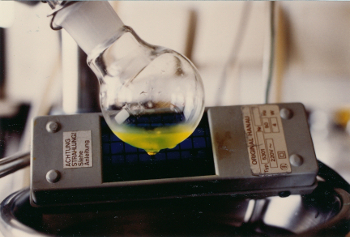
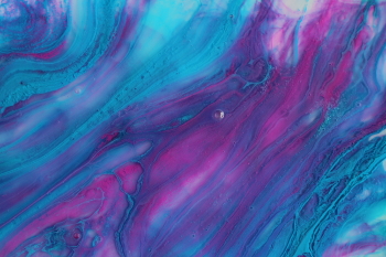
“Blue Vortex” Photo by Julia Bader Rapid stirring of a blue solution |
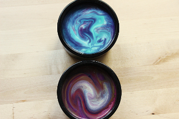
“Gluing Colors” Photo by Stefanie Neufeld-Busse Acrylic paint on a mixture of glues |
|||
|
|
||||
|
Entry 7 |
Entry 8 |
|||
|
|
 |
|||
|
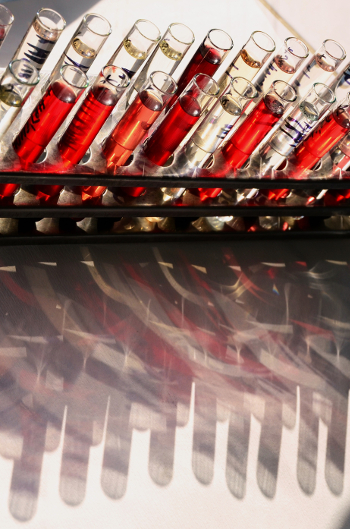
“Red Shadows” Photo by B. Vlachova and A. Novotna Rychtecka Biological samples for extraction |
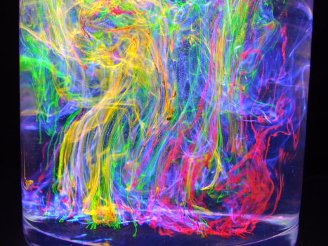
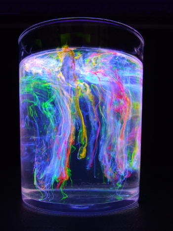
“Fluorescent Dyes 1” Photo by Bernard Valeur Fluorescent dyes slowly dissolving in a glycerol/ethanol mixture: fluorescein (yellow-green), rhodamine 101 (red), rhodamine 6G (orange), pyranine (blue), illumination by a UV-lamp |
|||
|
|
||||
|
Entry 9 |
Entry 10 |
|||
|
|
 |
|||
|
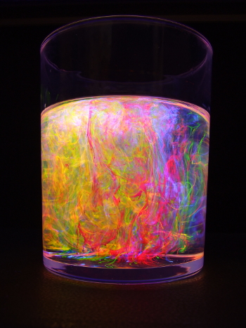
“Fluorescent Dyes 2”
Photo by Bernard Valeur Fluorescent dyes slowly dissolving in a glycerol/ethanol mixture: fluorescein (yellow-green), rhodamine 101 (red), rhodamine 6G (orange), pyranine (blue), illumination by a UV-lamp |
“Fluorescent Dyes 3”
Photo by Bernard Valeur Fluorescent dyes slowly dissolving in a glycerol/ethanol mixture: fluorescein (yellow-green), rhodamine 101 (red), rhodamine 6G (orange), pyranine (blue), illumination by a UV-lamp |
|||
|
|
||||
|
Entry 11 |
Entry 12 |
|||
|
|
 |
|||
|
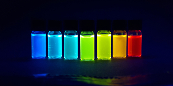
“Colorful Derivatization” Photo by Matthias Hempe Vials containing solutions of a series of photoluminescent emitter material derivatives for OLED applications |
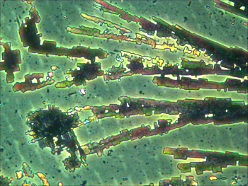
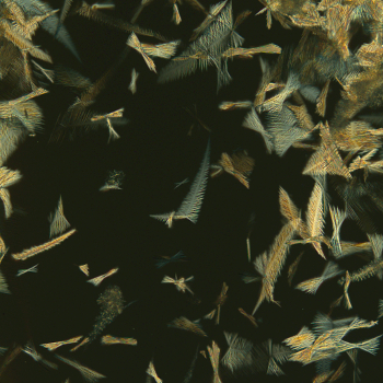
“Pillow Fight” Photo by Gregory York and Alfred Y. Lee A microscopic image of an organic cocrystal of a small molecule pharmaceutical compound; the ‘feather-like’ crystals were obtained via crystallization from the melt |
|||
|
|
||||
|
Entry 13 |
Entry 14 |
|||
|
|
|
|||
|
“Only yoU” Photo by Markus Zegke Highly air- and moisture-sensitive uranium(III) (blue) and uranium(IV) (green) compounds crystallizing side-by-side in an NMR tube, seen through a microscope |
“Fake Rainbow” Photo by Norbert Kemnitzer The mixed-up colors of the rainbow are accidentally obtained during the column chromatography of an unknown reaction product |
|||
|
|
||||
|
Entry 15 |
Entry 16 |
|||
|
|
|
|||
|
“Wide Range – which one to choose?” Photo by Norbert Kemnitzer A wide range of different colored bands are obtained during the column chromatography of a fluorescent dye; additional illumination by UV-light |
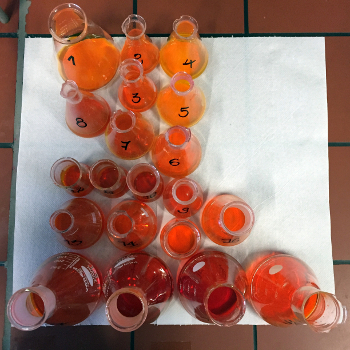
“Twenty Shades of Orange” Photo by Norbert Kemnitzer Fractions of a column chromatography |
|||
|
|
||||
|
Entry 17 |
Entry 18 |
|||
|
|
|
|||
|
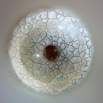
“Snow White” Photo by Norbert Kemnitzer Organic compound crystallizing in a fractal-like manner upon evaporation of the solvent; viewed through the neck of the round-bottom flask |
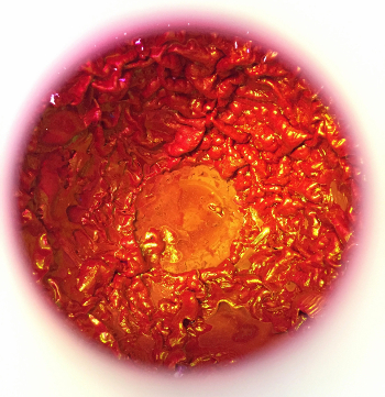
“Rose Red” Photo by Norbert Kemnitzer Organic dye-stuff after incomplete evaporation of the solvent; viewed through the neck of the round-bottom flask, it looks like an endoscopic insight into the digestive system |
|||
|
|
||||
|
Entry 19 |
Entry 20 |
|||
|
|
|
|||
|
“UV Argentum” Photo by Christian Schmitz Ag particles generated by a photochemical reaction under UV light |
“Near Infrared Polymers” Photo by Christian Schmitz Polymerization induced by near-infrared light and green-colored absorbers |
|||
|
|
||||
|
Entry 21 |
Entry 22 |
|||
|
|
|
|||
|
“Milky Way” Photo by ReAcTiON team Using colors to depict the dispersion of milk’s fatty acids due to its contact with dish soap, creating stellar-like surfaces |
“Glowing for Chemistry” Photo by Laurence Schmitz The calcite crystal represents inorganic chemistry; the super yellow represents organic chemistry; the bright blue aesculin of the chestnut branch represents biochemistry; all together they glow for the colorful world of chemistry! |
|||
|
|
||||
|
Entry 23 |
Entry 24 |
|||
|
|
|
|||
|
“Colorful Milk” Photo by Laurence Schmitz Milk with different indicators |
“Hot to cold?” Photo by Markus Plaumann Microscopic image after crystallization of an organic substrate |
|||
|
|
||||
|
Entry 25 |
Entry 26 |
|||
|
|
|
|||
|
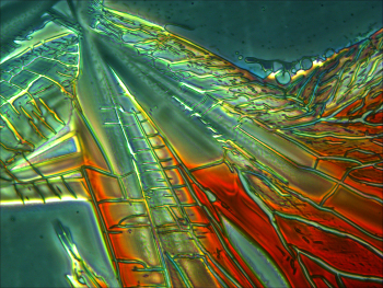
“Under Construction” Photo by Markus Plaumann Microscopic image after crystallization of an organic dye |
“Curtisin in Aceton” Photo by Martin Bröckelmann Dedicated to Wolfgang Steglich on the occasion of his 85th birthday (see also Eur. J. Org. Chem. 2004, 23, 4856–4863, https://doi.org/10.1002/ejoc.200400519) |
|||