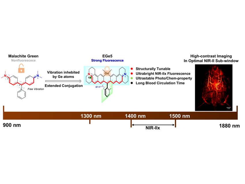
Near-infrared-II fluorescence imaging (900–1880 nanometers) offers unparalleled potential for high-resolution deep-tissue visualization, thanks to moderate tissue absorption, reduced photon scattering, and minimal background autofluorescence. By selectively absorbing photons, biological tissues suppress long-path scattered light, boosting the signal-to-noise ratio and enhancing image clarity. Within this spectrum, the NIR-IIx sub-window (1400–1500 nanometers) is especially promising, as it aligns with the absorption peak of water, the primary component of tissue. NIR-IIx imaging allows precise mapping of biological structures and dynamic processes, providing a powerful tool for disease diagnosis and treatment guidance.
Yet exploiting this window in vivo has remained challenging. The strict wavelength requirements and complex physiological environment demand small-molecule fluorophores with exceptional brightness and stability. Jun Qian, Zhejiang University, China, and colleagues have designed a xanthene-based fluorophore, EGe5, featuring germanium-incorporated extended conjugation to enhance both stability and fluorescence efficiency.
Their strategy involved three key design elements:
- Constructing a conjugated structure with a bis-naphthalene ring, which can effectively extend the conjugation length and result in a significant redshift of the fluorescence spectrum;
- Designing end groups as stronger electron-donating groups to enhance the intramolecular push–pull electronic effect and further shift the emission spectrum to longer wavelengths;
- Introducing germanium atoms, which can restrict the molecules free rotation, maintain a rigid-dye structure, and reduce non-radiative transitions, thereby enhancing fluorescence brightness.
The resulting EGe5 dyes emit around 946 nanometers in solution and 924 nanometers in serum, with fluorescence quantum yields of 10.5% and 3.3%, respectively.
Using the NIR-IIx window, the team achieved high-contrast imaging of intestinal wall vasculature in a mouse obstruction model. The remarkable clarity allowed precise delineation of diseased vessels, laying a solid foundation for accurate diagnosis.
This work represents the first demonstration of in vivo NIR-IIx imaging using an organic small-molecule dye. By enabling precise visualization of conditions such as intestinal and urinary tract obstructions, EGe5 opens new avenues for clinical imaging, with promising implications for diagnostics, therapy, and beyond.
- Germanium-Based Long-Conjugation Xanthene Fluorophores for Bioimaging in Optimal NIR-II Sub-Window.
Jin Li, Qiming Xia, Jiayi Li, Xiaoming Yu, Zhe Feng, Yuhuang Zhang, Tianxiang Wu, Zhongmin Xu, Hui Lin, Jun Qian
Aggregate 2025
https://doi.org/10.1002/agt2.70148