Lysosomes are essential cellular organelles responsible for degrading biomolecules and clearing damaged organelles. Although super-resolution imaging technologies have revolutionized subcellular visualization beyond the limits of conventional fluorescence imaging, their broader application is still hindered by fluorescent probes that often require complex synthesis and show poor intracellular stability, low photostability, and limited near-infrared (NIR) imaging capabilities. Moreover, interpreting complex imaging data into coherent and concise textual conclusions remains a difficult, time-consuming, and error-prone task, often requiring expertise across multiple disciplines.
To address these challenges, Yujie Sun, Jiajie Diao (University of Cincinnati, USA), and colleagues developed PA-2, a quinolinium-based fluorescent probe tailored for NIR super-resolution imaging. PA-2 forms nanoaggregates in aqueous solution, enters cells via energy-dependent endocytosis, and localizes specifically in lysosomes. Using PA-2, the researchers visualized dynamic lysosomal processes—including autophagy, mitochondrial–lysosome contacts, and mitophagy—in living cells with high clarity and resolution.
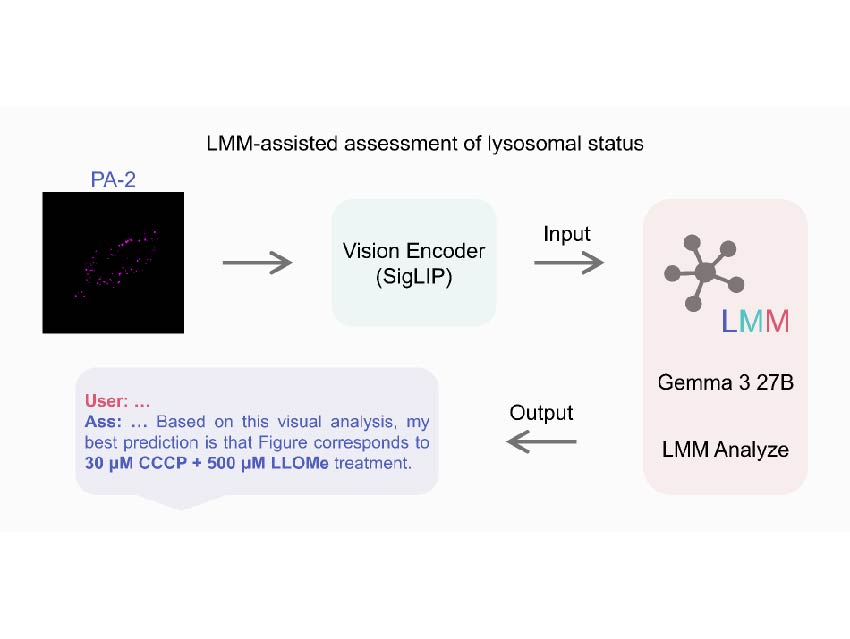
To overcome limitations in image interpretation, this work further integrated imaging with a Large Multimodal Model (LMM, an artificial intelligence tool) through using Google’s Gemma 3 27B model with SigLIP vision encoder. The LMM was trained to interpret super-resolution images of PA-2, distinguish lysosomal status under various drug treatments (e.g., CCCP and LLOMe; commonly used chemical tools in cell biology to manipulate lysosomal and mitochondrial function), and generate accurate and explainable conclusions. Remarkably, the LMM outperformed human evaluators in image analysis, enabling faster and more reliable assessments of lysosomal status.
This work highlights the strong potential of combining advanced fluorescent probe design with AI-assisted image interpretation to drive innovation in bioimaging. The integration of chemical tools and AI offers a transformative path toward faster, accurate, and scalable discoveries in biological research and beyond.
- A Versatile Near-Infrared Fluorescent Probe for Fast Assessment of Lysosomal Status via a Large Multimodal Model
Rui Chen, Eugene Lee, Yuxin Wang, Aditya Yadav, Minling Zhong, Pragti, Yujie Sun, Jiajie Diao
Aggregate 2025
https://doi.org/10.1002/agt2.70118