Observing Metabolites Directly
Living cells can react to disturbances with a changed metabolism, but direct observation of trafficking metabolites in live cells is difficult. Marc Vendrell, The University of Edinburgh, UK, and colleagues have developed a class of remarkably small fluorophores called SCOTfluors.The dyes emit light in the visible to near-infrared range and can be attached to common metabolites.
When a living cell changes its metabolism, because of an external signal or because there is something wrong with it, the trafficking of metabolites will change. As metabolites are usually small molecules, typically cells have to be destroyed and the metabolome extracted in order to record such changes. Alternatively, the metabolites could be labeled with a dye, which reveals itself through a fluorescence signal under a microscope.
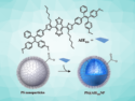
However, common dyes are often much larger molecules than the metabolite to be labeled. The team set out to develop the smallest fluorophores to date, which can be attached to typical metabolites such as lipids, sugars, and carboxylic acids.
Small and Tunable Fluorophores
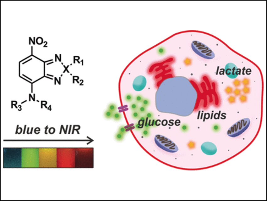
Fluorescent dyes usually contain fused aromatic rings, which provide a conjugated electronic system, the chromophore. To minimize the size of the dyes, the scientists worked with nitrobenzodiazoles, which contain only one benzene ring, an electronically active nitro group, and a fused diazo ring. This structure proved to be beneficial in two ways: first, it is really small compared to the other fluorescent dyes, and second, the scientists could tune the emission wavelengths just by changing one atom in the molecule; for example, by replacing an oxygen atom with nitrogen, sulfur, selenium, or carbon.
To check whether metabolite tracking was possible by fluorescence labeling, the scientists attached fluorophores either to ceramide, which is a member of the sphingolipid class, glucose, or lactic acid. Then, they added the labeled metabolites to cultures of human cells and the metabolites could be localized in the respective organelles. It was even possible to associate the recycling rates of lactate in hypoxic or normoxic cells—cells that contain different levels of oxygen, as cancer cells do. They also fed labeled glucose to zebrafish embryos and monitored its uptake into their developing brains.
The team pointed out that the tunability of their mini-fluorophores, which they named SCOTfluors, could be another advantage. They prepared different fluorophores using the same molecular platform and using similar synthetic steps. Not only could they label different metabolites with different colors, but they were also able to follow their uptake profiles in cancer cells simultaneously. This work gives an example of how new, small fluorophores shed light into the fascinating metabolic machinery of living cells.
- SCOTfluors: Small, Conjugatable, Orthogonal, and Tunable Fluorophores for In Vivo Imaging of Cell Metabolism,
Sam Benson, Antonio Fernandez, Nicole D. Barth, Fabio de Moliner, Mathew H. Horrocks, C. Simon Herrington, Jose Luis Abad, Antonio Delgado, Lisa Kelly, Ziyuan Chang, Yi Feng, Miyako Nishiura, Yuichiro Hori, Kazuya Kikuchi, Marc Vendrell,
Angew. Chem. Int. Ed. 2019.
https://doi.org/10.1002/anie.201900465