Optical microscopy provides a non-invasive way to study living systems. The resolution of this technique, however, is limited by the diffraction of light. Single-molecule localization microscopy overcomes this limit by using probes that can be transformed between fluorescent and non-fluorescent forms. These simple on/off switches can be bound to proteins and enable the observation of these proteins based on their expression levels.
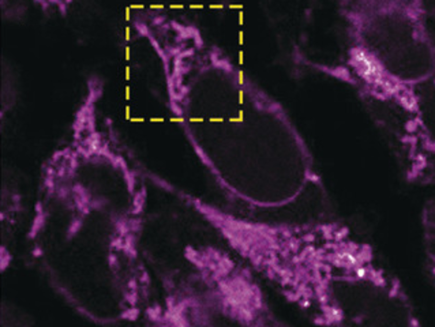
Zacharias Thiel and Pablo Rivera-Fuentes at ETH Zurich, Switzerland, developed a probe that enables the observation of single enzymes based on their activity. Nitroreductases, enzymes involved in the activation of cancer drugs, were used for this approach. The researchers designed a diazoindanone derivative of a rhodamine dye. This probe was activated sequentially by nitroreductases and light to generate a fluorophore. This fluorophore is crosslinked to the enzyme, which limits its diffusion and makes the imaging more precise.
Using this probe, the intracellular localization of active nitroreductases was mapped with single-molecule precision in a living cell. Such information could be essential for the development of improved therapies with fewer side effects.
- Single-Molecule Imaging of Active Mitochondrial Nitroreductases Using a Photo-Crosslinking Fluorescent Sensor,
Zacharias Thiel, Pablo Rivera-Fuentes,
Angew. Chem. Int. Ed. 2019.
https://doi.org/10.1002/anie.201904700