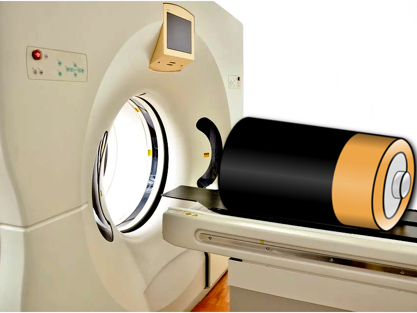
X-ray computed tomography has become a pivotal technique used by archeologists and materials scientists to unveil what is inside compact bodies without destroying them. Researchers have adapted this technique to commercial lithium batteries.
In combination with neutron tomography and by applying a virtual “unwinding” technology, they obtained a telling picture of the actual morphological changes when the cell was discharging. The tomography showed uneven lithiation and regions of significant electrode cracking. Therefore, the researchers recommend such analyses for battery optimization.
Lithium—An Elusive Element for X-Rays
Although the electrode processes in lithium batteries have been well established, the actual events and the morphological impacts are difficult to observe in an operating battery cell. Usually, batteries must be dismantled mechanically to detect cracks, material failures, and other features.
One state-of-the-art technique to see through massive bodies is X-ray tomography, which is the slice-wise spectroscopic scanning and computed three-dimensional reconstruction of artifacts – similar to the magnetic resonance imaging (MRI) technique in medicine, but using X-rays or other radiation, not magnetic resonance.
Probably the most spectacular results in this field come from the field of archeology where, for example, any detail of a mummy inside a locked sarcophagus can be revealed or the finer mechanical features of a precious ancient tool can be brought to light. Less appealing, but no less useful, is the scanning of massive materials such as construction items to identify internal cracks and defects.
However, as X-rays interact with electrons, X-ray tomography only works well for heavy elements with large electron clouds, which give sufficient contrast. Lithium, as one of the lightest elements, fails to produce sharp features in X-ray scans. This means that in lithium-ion batteries, only the cathode, which is made of transition metal oxides, is visible. Thus, although X-ray tomography can unveil morphological changes in the cathode, the processes which lead to these changes are not seen.
Combination and Computing
Neutron tomography, however, reveals lithium. Neutrons interact with nuclei and are more sensitive for light elements, the nuclei of which tend to be more closely packed. In a large international collaboration including teams from University College London, the Harwell Science and Innovation Campus, Harwell, UK, the Université Grenoble Alpes, France, the National Renewable Energy Laboratory, Golden, USA, and the Technische Universität Berlin, Germany, researchers investigated commercial Duracell lithium CR2 primary cells using both techniques, X-ray tomography at a synchrotron facility and neutron tomography.
The rechargeable Duracell batteries consist of a pure lithium foil as the anode and a cathode made of MnO2 with an interwoven nickel mesh as the current collector. The cathode, electrolyte (plus separator), and anode are tightly rolled up to make the typical cylindrical battery form. The scientists analyzed one such cell at the European Synchrotron Radiation Facility (ESRF) beamline in Grenoble and another one at the neutron imaging beamline at the Helmholtz Centre Berlin. Both cells were first in the charged state, then in two partially discharged states.
After the computed reconstruction, both tomography techniques provided a complementary and remarkably detailed view of the three-dimensional electrochemical processes occurring in the battery from top to bottom and from the inner electrolyte reservoir to the utmost winding. To reduce complexity and exclude the strongly absorbing nickel mesh from the results, the scientists also “unrolled” the electrodes by a special computing technique.
Cracks and Electrolyte Consumption
From the wealth of information gained, the scientists reported two main results: first, that it was indeed possible to observe the morphological changes in both electrodes and, second, these changes were distributed surprisingly unevenly throughout the cell. It appeared that at positions where compression was highest, as in the middle section of the battery cell and in the inner windings, less lithium found its way into the cathode, which also expanded less than in the outer windings.
The scientists also observed cracks from manufacturing, especially at the sites of the nickel mesh, which grew and led to delamination when the cathode was lithiated. These cracks were quickly filled with excess electrolyte, which, at the same time, disappeared from the original reservoir in the center of the cell.
The analysis provided a fascinating visualization of what happens to the materials in a practical operating battery. However, even greater levels of detail are desirable. Although the neutron tomography was shown to complement the X-ray tomography, the researchers reported that the electrolyte was still not clearly discernable from the lithium meaning that they could not determine the charging state from the lithium consumption. Further refinement of the techniques and more elaborate computation is needed to overcome these limitations.
- 4D imaging of lithium-batteries using correlative neutron and X-ray tomography with a virtual unrolling technique,
Ralf F. Ziesche, Tobias Arlt, Donal P. Finegan, Thomas M. M. Heenan, Alessandro Tengattini, Daniel Baum, Nikolay Kardjilov, Henning Markötter, Ingo Manke, Winfried Kockelmann, Dan J. L. Brett, Paul R. Shearing,
Nat. Commun. 2020.
https://doi.org/10.1038/s41467-019-13943-3
![]()