Modeling Melanopsin to Understand the Circadian Rhythm
The term “biological clock” has become familiar to many people, the notion of our daily lives being governed not by sunrise and sunset but by some internal, circadian, rhythm. International travelers, shift-workers, and parents of new-born children are often well aware of the detrimental effects on our well-being when the mainspring of our biological clock is over wound. Now, insights into the chemistry that underpins the rhythm of our lives, and indeed that of all mammals, have come to light.
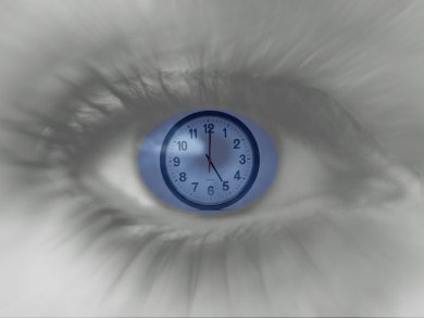
Writing in the Journal of the American Chemical Society, chemists at Yale University, New Haven, Connecticut, USA, have reported details of the active site in the pigment molecule melanopsin. Melanopsin is present in the eye, but is not involved in vision. Instead it responds to light that enters the eye and reaches neurones (ganglions) deep within the retina. The pigment absorbs blue light, which hints at why there are so many current concerns about the effects of ubiquitous electric lighting and computer and mobile device screens that are always in our field of view. Absorption in this wavelength stimulates the pigment and sends a neuronal signal to the suprachiasmatic nuclei. This is a small region of the brain known to be at the heart of regulating the circadian rhythms of our neurones and hormones and matching them to the natural cycle of the 24-hour day.
While melanopsin is now well known to those working in the field, its structure is yet to be pinned down by X-ray crystallography or spectroscopy, or indeed by any other method. Sivakumar Sekharan, Jennifer Wei, and Victor Batista at Yale, have now shed some light on this problem by proposing a model for the active site of the pigment found in the mouse. The model is based on earlier crystal structure studies of the closely related squid visual rhodopsin pigment. Hints from these studies and those on bovine rhodopsin suggest that a single mutation between the gene for visual pigments like rhodopsin and non-visual melanopsin, is responsible for the switch between light sensors needed for sight and the photoreceptors involved in other, non-visual processes, such as setting the biological clock.
It is worth mentioning that melanopsin and its chemical cousins are all “G-protein-coupled receptors”, GPCRs, which have, of course, come to the fore recently as being the focus of this year’s Nobel Prize for Chemistry awarded to Robert Lefkowitz of the Howard Hughes Medical Institute and Brian Kobilka of the Stanford University School of Medicine, both USA. As such, there is much new attention on the biochemistry of signal transmission involving GPCRs. Parallel work will undoubtedly help inform structural and modeling studies of melanopsin, rhodopsin, and their ilk.
Validity of Model
According to Sekharan and colleagues melanopsin in primates and mice controls visual processing and is thought to optimize the visual pathways depending on the time of day. The phrases “early morning” and “bleary eyed” often go together for many people and hint at the underlying connection between time of day, one’s biological clock, and the dependence of visual acuity on such characteristics. Melanopsin resembles the photoreceptor pigments of higher invertebrates, such cephalopods and arthropods, based on its primary protein sequence and the photoactivation cascade to which it succumbs when light shines on it.
The team’s model based on the squid rhodopsin crystal structure is thus perfectly valid. The researchers created a QM/MM model using the two-layer ONIOM scheme – which models large molecules by defining two or three layers within the structure that are treated at different levels of accuracy – with electronic-embedding (EE) implemented in the program Gaussian09. Squid rhodopsin has an absorption maximum at a wavelength of 490 nm, while the model reveals it to be 447 nm in melanopsin, which the team says is consistent with observed photoreceptor absorbance. The 43 nm shift can be explained by an increase in bond-length alternation due to the single mutation that differentiates melanopsin from rhodopsin.
“Understanding the structure, wavelength sensitivity, and spectral tuning of melanopsin is the first step toward manipulating the regulation of circadian rhythms,” the researchers say. “As light is a powerful regulator of the circadian system, the findings could allow us to optimize the use of light in therapeutic applications,” they conclude.
Alexander Lerchl of Jacobs University in Bremen, Germany, has worked on the structure of melanopsin and predicted a plausible structure back in 2005 published in the journal Neuroscience Letters. “The [new] study is interesting because it helps us to understand the absorption characteristics with a peak in the blue part of the spectrum,” he told ChemViews magazine, blue being the important part of the spectrum associated with body clock response it seems.
- The Active Site of Melanopsin: The Biological Clock Photoreceptor,
Sivakumar Sekharan, Jennifer N. Wei, Victor S. Batista,
J. Am. Chem. Soc. 2012.
DOI: 10.1021/ja308763b
Also of interest:
- Towards Understanding Color Perception
A rhodopsin protein mimic has been designed where wavelength absorption can be drastically tuned - The Eye of the Beholder
New calculations explain why vision uses the 11-cis form of the pigment retinal, rather than any of the other possible forms