The Nobel Prize in Chemistry 2014 has been awarded jointly to Eric Betzig, Howard Hughes Medical Institute, Ashburn, VA, USA, Stefan W. Hell, Max Planck Institute for Biophysical Chemistry, Göttingen, and German Cancer Research Center, Heidelberg, Germany, and William E. Moerner, Stanford University, CA, USA, “for the development of super-resolved fluorescence microscopy”.
Their work has brought optical microscopy into the nanodimension. With the help of fluorescent molecules they circumvented the long presumed limitation of optical microscopy that it would never obtain a better resolution than half the wavelength of light.
Stefan Hell developed the stimulated emission depletion (STED) microscopy method: One laser beam stimulates fluorescent molecules to glow, a second laser beam cancels out all fluorescence except for that in a nanometre-sized volume.
Eric Betzig and William Moerner, working separately, used the possibility to turn the fluorescence of individual molecules on and off for their single-molecule microscopy method: the same area is imaged multiple times, so that only a few interspersed molecules glow each time. Superimposing these images yields a dense super-image resolved at the nanolevel.
Eric Betzig, born 1960 in Ann Arbor, MI, USA, gained his Ph.D. from Cornell University, Ithaca, NY, USA, in 1988. He then worked at AT&T Bell Laboratories, USA, in the Semiconductor Physics Research Department. In 1996, Betzig became Vice President of research and development at Ann Arbor Machine Company, then owned by his father.
Currently Betzig is Group Leader at Janelia Farm Research Campus, Howard Hughes Medical Institute, Ashburn, VA, USA.
Stefan W. Hell, born 1962 in Arad, Romania, received his Ph.D. from the University of Heidelberg, Germany, in 1990. From 1991 to 1993 he worked at the European Molecular Biology Laboratory, Heidelberg, from 1993 to 1996 he worked as a group leader at the University of Turku, Finland, and from 1993 to 1994 he was for six months a visiting scientist at the University of Oxford, UK. Hell received his habilitation in physics from the University of Heidelberg in 1996 and became a director of the Max Planck Institute for Biophysical Chemistry in 2002; he established the department of Nanobiophotonics there. Since 2003 Hell has also been the leader of the department Optical Nanoscopy division at the German Cancer Research Center (DKFZ), Heidelberg.
Currently Hell is Director at the Max Planck Institute for Biophysical Chemistry, Göttingen, and Division Head at the German Cancer Research Center, Heidelberg, Germany.
William E. Moerner, born 1953 in Pleasanton, CA, USA, gained his Ph.D. from Cornell University, Ithaca, NY, USA, in 1982. From 1981 to1995, he worked at the IBM Almaden Research Center, San Jose, CA, USA. After an appointment as Visiting Guest Professor of Physical Chemistry at ETH Zurich, Switzerland, from 1993 to 1994, Moerner assumed the Distinguished Chair in Physical Chemistry in the Department of Chemistry and Biochemistry at the University of California, San Diego, USA, from 1995 to 1998. In 1998 his research group moved to Stanford University where he became Professor of Chemistry, Harry S. Mosher Professor in 2003.
Currently, Moerner is Harry S. Mosher Professor in Chemistry and Professor, by courtesy, of Applied Physics at Stanford University, Stanford, CA, USA.
Moerner was one of the guest editors of the special issue of ChemPhysChem on Superresolution Imaging earlier this year which included an editorial by himself and an open access contribution by the group of Hell. (the issue is free-to-read for a limited time)
Publications of Eric Betzig
- K. Wang, D. E. Milkie, A. Saxena, P. Engerer, T. Misgeld, M. E. Bronner, J. Mumm, E. Betzig, Rapid adaptive optical recovery of optimal resolution over large volumes, Nat. Methods 2014,11(6), 625–8. DOI: 10.1038/nmeth.2925
- L. Gao, L. Shao, B. C. Chen, E. Betzig, 3D live fluorescence imaging of cellular dynamics using Bessel beam plane illumination microscopy, Nat. Protoc. 2014, 9(5),1083–101. DOI: 10.1038/nprot.2014.087
- E. Betzig, G. H. Patterson, R. Sougrat, O. W. Lindwasser, S. Olenych, J. S. Bonifacino, M. W. Davidson, J. Lippincott-Schwartz, H. F. Hess, Imaging intracellular fluorescent proteins at nanometer resolution, Science 2006, 313 (5793), 1642–1645 . DOI: 10.1126/science.1127344
- Eric Betzig, Proposed method for molecular optical imaging, Opt Lett. 1995, 20 (3), 237–239. DOI: 10.1364/OL.20.000237
Publications of Stefan W. Hell
- S. Arroyo-Camejo, A. Lazariev, S. W. Hell, G. Balasubramanian, Room temperature high-fidelity holonomic singe-qubit gate on a solid-state spin, Nature Commun. 2014, 5, 1–5. DOI: 10.1038/ncomms5870
- G. Lukinavicius, L. Reymond, E. D’Este, A. Masharina, F. Göttfert, H. Ta, A. Güther, M. Fournier, S. Rizzo, H. Waldmann, C. Blaukopf, C. Sommer, D. W. Gerlich, H. Arndt, S. W. Hell, K. Johnsson, Fluorogenic probes for live-cell imaging of the cytoskeleton, Nature Meth. 2014. DOI: 10.1038/nmeth.2972
- S. K. Saka, A. Honigmann, C. Eggeling, S. W. Hell, T. Lang, S. Rizzoli, Multi-protein assemblies underlie the mesoscale organization of the plasma membrane, Nature Commun. 20014, 5, 1–14. DOI: 10.1038/ncomms5509
- F. Görlitz, P. Hoyer, H. J. Falk, L. Kastrup, J. Engelhardt, S. W. Hell, A STED Microscope Designed for Routine Biomedical Applications, Prog. Electromagn. Res. 2014, 147, 57–68.
- K. I. Willig, H. Steffens, C. Gregor, A. Herholt, M. J. Rossner, S. W. Hell, Nanoscopy of Filamentous Actin in Cortical Dendrites of a Living Mouse, Biophys. J. 2014, 106, L01–L03. DOI: 10.1016/j.bpj.2013.11.1119
 Flavie Lavoie-Cardinal, Nickels A. Jensen, Volker Westphal, Andre C. Stiel, Andriy Chmyrov, Jakob Bierwagen, Ilaria Testa,
Stefan Jakobs, Stefan W. Hell, Two-Color RESOLFT Nanoscopy with Green and Red Fluorescent Photochromic Proteins, ChemPhysChem 2014. DOI: 10.1002/cphc.201301016- Jensen, N. A., J. G. Danzl, K. I. Willig, F. Lavoie-Cardinal, T. Brakemann, S. W. Hell, S. Jakobs, Coordinate-Targeted and Coordinate-Stochastic Super- Resolution Microscopy with the Reversibly Switchable Fluorescent Protein Dreiklang, ChemPhysChem 2014, 15, 756-762. DOI: 10.1002/cphc.201301034
- S. J. Sahl, M. Leutenegger, S. W. Hell, C. Eggeling, High-Resolution Tracking of Single-Molecule Diffusion in Membranes by Confocalized and Spatially Differentiated Fluorescence Photon Stream Recording, ChemPhysChem 2014, 15, 771-783. DOI: 10.1002/cphc.201301090
- K. Kolmakov, C. A. Wurm, D. N. H. Meineke, F. Göttfert, V. P. Boyarskiy, V. N. Belov, S. W. Hell, Polar Red-Emitting Rhodamine Dyes with Reactive Groups: Synthesis, Photophysical Properties, and Two-Color STED Nanoscopy Applications, Chem. Eur. J. 2014, 20, 146-157. DOI: 10.1002/chem.201303433
- A. B. Wong, M. A. Rutherford, M. Gabrielaitis, T. Pangrsic, F. Göttfert, T. Frank, S. Michanski, S. Hell, F. Wolf, C. Wichmann, T. Moser, Developmental refinement of hair cell synapses tightens the coupling of Ca2+ influx to exocytosis, EMBO J. 33, 247-264. DOI: 10.1002/embj.201387110
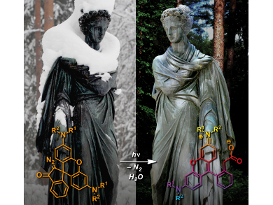
- Vladimir N. Belov, Gyuzel Yu. Mitronova, Mariano L. Bossi, Vadim P. Boyarskiy, Elke Hebisch, Claudia Geisler, Kirill Kolmakov, Christian A. Wurm, Katrin I. Willig, Stefan W. Hell, Masked Rhodamine Dyes of Five Principal Colors Revealed by Photolysis of a 2-Diazo-1-Indanone Caging Group: Synthesis, Photophysics, and Light Microscopy Applications, Chem. Eur. J. 2014. DOI: 10.1002/chem.201403316
News on this article published in ChemViews Mag.: Hidden Colors by Anne Deveson
- S. W. Hell, M. Kroug, Ground-state-depletion fluorscence microscopy: A concept for breaking the diffraction resolution limit, Appl. Phys. B 1995, 60 (5), 495–497. DOI:
- S. W. Hell, J. Wichmann, Breaking the diffraction resolution limit by stimulated emission: stimulated-emission-depletion fluorescence microscopy, Opt Lett. 1994, 19(11), 780–782. DOI: 10.1364/OL.19.000780
Publications of William E. Moerner
- Matthew D. Lew, W. E. Moerner, Azimuthal polarization filtering for accurate, precise, and robust single-molecule localization microscopy, Nano Letters 2014. DOI: 10.1021/nl502914k
- Marissa K. Lee, Prabin Rai, Jarrod Williams, Robert J. Twieg, W. E. Moerner, Small-Molecule Labeling of Live Cell Surfaces for Three-Dimensional Super-Resolution Microscopy, J. Amer. Chem. Soc. 2014. DOI: 10.1021/ja508028h
- Yoav Shechtman, Steffen J. Sahl, Adam S. Backer, W. E. Moerner, Optimal Point Spread Function Design for 3D Imaging, Phys. Rev. Lett. 2014, 113, 133902. DOI: 10.1103/PhysRevLett.113.133902
- Yin Loon Lee, Joshua Santé, Colin J. Comerci, Benjamin Cyge, Luis F. Menezes, Feng-Qian Li, Gregory G. Germino, W. E. Moerner, Ken-Ichi Takemaru, and Tim Stearns, Cby1 promotes Ahi1 recruitment to a ring-shaped domain at the centriole–cilium interface and facilitates proper cilium formation and function, Mol. Biol. Cell 2014, 25 (19), 2919–2933. DOI: 10.1091/mbc.E14-02-0735
- Julie S. Biteen, Erin D. Goley, Lucy Shapiro, W. E. Moerner, Three-Dimensional Super-Resolution Imaging of the Midplane Protein FtsZ in Live Caulobacter crescentus Cells Using Astigmatism, ChemPhysChem 2012, 13 (4), 1007–1012. DOI: 10.1002/cphc.201100686
- Randall H. Goldsmith, W. E. Moerner, Watching conformational- and photodynamics of single fluorescent proteins in solution, Nature Chem. 2010, 2, 179. DOI: 10.1038/nchem.545
- Lee, Jaeckel, Moerner, Bao, Micrometer-Sized DNA-Single Fluorophore-DNA Supramolecule: Synthesis and Single-Molecule Characterization, Small 2009, 5 (21), 2418. DOI: 10.1002/smll.200900494
- Robert M. Dickson, Andrew B. Cubitt, Roger Y. Tsien, W. E. Moerner, On/off blinking and switching behaviour of single molecules of green fluorescent protein, Nature 1997, 388, 355–358.
- W. E. Moerner, L. Kador, Optical detection and spectroscopy of single molecules in a solid, Phys. Rev. Lett. 1989, 62, 2535. DOI: 10.1103/PhysRevLett.62.2535
Also of interest
- Who’s Next? Nobel Prize in Chemistry 2014
In this prediction, Stefan Hell and William Moerner received two votes each - Hidden Colors by Anne Deveson
Caged rhodamine dyes used as “hidden” markers in subdiffractional and conventional light microscopy

- Video: Lecture by S. Hell On Super-Resolution: Overview and Stimulated Emission Depletion Microscopy
- Press release: Wiley Authors Awarded 2014 Nobel Prize in Chemistry
- Microscopy Research & Technique Stefan W. Hell Virtual Issue; articles are freely available until the end of 2014


