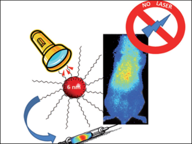
Fluorescent nanoparticles (NPs) are of interest for optical imaging in medical diagnosis, but their fluorescence signal is often obscured by tissue autofluorescence, which arises from the continuous in situ excitation needed to make the NPs fluoresce. Persistent luminescence nanoparticles (PLNPs) allow very sensitive optical detection in vivo by avoiding the autofluorescence of tissues. However, because of their large hydrodynamic size (> 100 nm), PLNPs are quickly taken up and sequestered by the reticuloendothelial system (RES).
Yoann Lalatonne, Université Paris 13, France, and co-workers have created ultra-small ZnGa2O4:Cr3+ nanoparticles (6 nm in diameter) that exhibit near-infrared persistent luminescence and high stability under physiological conditions. The PLNPs were synthesized by a non-aqueous sol-gel method assisted by microwave irradiation, followed by coating with polyethylene glycol (PEG) phosphonate moieties as surface ligands.
The PLNPs were excited prior to injection into mice and the resultant persistent luminescence signal was detected for up to four hours after excitation, without the need for excitation throughout the imaging. Ex vivo analysis showed retention of the particles in various organs, including the heart, lungs, liver, spleen, and kidneys. The small nanoparticle size and PEG passivation reduce the RES clearance, enhance the blood circulation time, and allow widespread organ distribution, which means that these nanoparticles could be used for a variety of imaging targets such as those for cancer or cardiovascular diseases. The minimal RES capture may also prevent their long term accumulation and possible toxicity concerns.
- Non-Aqueous Sol-Gel Synthesis of Ultra Small Persistent Luminescence Nanoparticles for Near-Infrared In Vivo Imaging,
Eliott Teston, Sophie Richard, Thomas Maldiney, Nicole Lièvre, Guillaume Yangshu Wang, Laurence Motte, Cyrille Richard, Yoann Lalatonne,
Chem. Eur. J. 2015.
DOI: 10.1002/chem.201406599