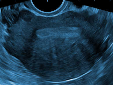
Medical ultrasound imaging is an essential diagnostic tool and is often used as a complementary technique to more expensive or invasive imaging techniques. Perfluorocarbons are commonly used as ultrasound contrast agents because they undergo a large pressure change when they are stimulated by ultrasonic radiation and form gaseous microbubbles, thus providing a contrast under different ultrasound modes.
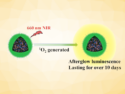
Jeffrey Reinhart, University of California, San Diego, La Jolla, USA, and colleagues have developed an ultrasound contrast agent by loading polydopamine nanoparticles in a size range of 70–50 nm with perfluoropentane. The polydopamine nanoparticles were synthesized using wet chemical methods. The nanoparticles were then loaded with the perfluoropentane molecules using a combination of immersion, freeze-drying, and ultrasonication dispersion methods.
The loaded nanoparticles show a strong and persistent ultrasound contrast in both aqueous suspensions and ex-vivo tissue samples. The contrast under continuous imaging by color Doppler ultrasound was found to persist for up to one hour when using particles with a size of 135 nm. Particles with a diameter of 170 nm were found to provide the best signal-to-noise ratio. The synthetic process is tunable and similar contrast agents could be used for other imaging approaches in the future.
- Perfluorocarbon-loaded polydopamine nanoparticles as ultrasound contrast agents,
Yijun Xie, James Wang, Zhao Wang, Kelsey A. Krug, Jeffrey D. Rinehart,
Nanoscale 2018, 10, 12813–12819.
https://doi.org/10.1039/c8nr02605j