Atomic force microscopy (AFM) is showing promise as a probe of cancer cell morphology and stress. It could be used to help pathologists assess suspicious cells from a biopsy and detect cancer at an early stage. It might also be used to measure the effects of cancer therapeutics on cells.
AFM probes the elasticity of materials by measuring the resistive force the material exerts on a sharp or spherical tip on the end of a flexible cantilever as it is pressed into the sample. When studying cancer cells, a plot of resistance versus the probe’s depth yields a force curve that provides information about the cell’s stiffness.
James Gimzewski and colleagues, University of California – Los Angeles, USA, have used this to determine and quantitate the elasticity differences between metastatic and benign cells [1]. They found that lung, breast, and pancreatic metastatic cells were, on average, 70 % softer than benign cells and the distribution of cell stiffness values for normal cells was much broader than for diseased cells. This indicates that diseased cells are more morphologically similar to one another and implies that the softness aids in the spreading of the metastatic cells.
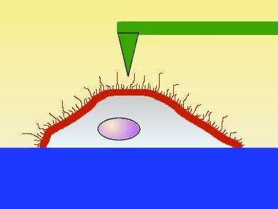
In addition to cancer cells, Robert Ross and co-workers, Arizona State University, USA, have shown that precancerous cells show signs of softness [2], while Igor Sokolov and co-workers, Clarkson University, New York, USA, found that membrane protrusions on the outside of cells are different on the surface of normal and cancerous cells [3]. They showed that cancerous cells have a brush of long, sparse whiskers (microvilli, see picture) interspersed with short, dense whiskers, whereas normal cells have a dense coating of whiskers that are all one length.
As more discoveries about cancer cells are made with AFM, the need for standardized methods and protocols grows. Gimzewski and his group are trying to set up a lab in the cytology department at UCLA’s Medical Center in order to begin automating the process. There, they will look at cells both optically, as pathologists do, and with AFM in order to create a useful tool for clinicians. Gimzewski also has cofounded a company, Edeixa, to commercialize the technology.
- Using The Force On Cancer,
Lauren K. Wolf,
C&ENews 2011, 89 (21), 34–36. (May 23)
References
- Sarah E. Cross, Yu-Sheng Jin, Jianyu Rao, James K. Gimzewski, Nanomechanical analysis of cells from cancer patients, Nat. Nanotechnol. 2007, 2, 780-783. DOI: 10.1038/nnano.2007.388
- A. Fuhrmann, J. R. Staunton, V. Nandakumar, N. Banyai, P. C. W. Davies, R. Ros, Phys. Biol., AFM stiffness nanotomography of normal, metaplastic and dysplastic human esophageal cells, Phys. Biol. 2011, 8. DOI: 10.1088/1478-3975/8/1/015007
- S. Iyer, R. M. Gaikwad, V. Subba-Rao, C. D. Woodworth, Igor Sokolov, Atomic force microscopy detects differences in the surface brush of normal and cancerous cells, Nat. Nanotechnol. 2009, 4, 389–393. DOI: 10.1038/nnano.2009.77