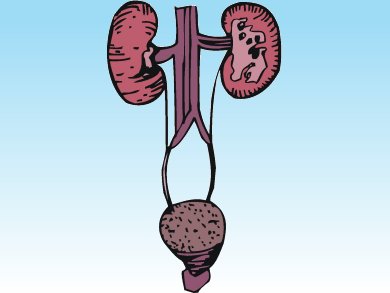
The death of the cells forming the tubules that transport urine to the ureters causes acute tubular necrosis. Diagnostic tools allowing an early detection of this disease and its differentiation from acute glomerulonephritis are essential. Nevertheless, they are currently lacking.
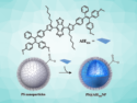
Menna R. Clatworthy and colleagues, University of Cambridge, UK, demonstrated that necrotic cells’ ability to uptake hyperpolarized [1,4-13C2]fumarate and convert it to [1,4-13C2]malate can constitute a new diagnostic strategy.
Using mice models, the scientists demonstrated that magnetic resonance imaging (MRI) of hyperpolarized [1,4-13C2]fumarate’s metabolism can identify renal acute tubular necrosis in its early phase, when histological changes are minor. Moreover, since in acute glomerulonephritis fumarate is not converted to malate, this technique provides a differential diagnosis between the two diseases.
- Magnetic resonance imaging with hyperpolarized [1,4-13C2]fumarate allows detection of early renal acute tubular necrosis,
M. R. Clatworthy, M. I. Kettunen, D. E. Hu, R. J. Mathews, T. H. Witney, B. W. Kennedy, S. E. Bohndiek, F. A. Gallagher, L. B. Jarvis, K. G. Smith, K. M. Brindle,
Proc. Natl. Acad. Sci. 2012.
DOI:10.1073/pnas.1205539109