Microscopic gas-filled spheres of silica can mark the location of early-stage tumors to show their position by using ultrasound imaging in the operating room.
A team of chemists, radiologists and surgeons lead by William Trogler, University of California, San Diego, USA, created spheres of silica filled with perfluoropentane gas. The microbubbles can be injected into clusters of abnormal cells using a thin needle. The balls have high stability and adhere to breast tissue, allowing radiologists to inject them days before surgery.
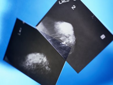
Ultrasound pressure waves burst the microbubbles. The gas is expelled on breakage of the ball, creating an efficient contrast agent for color Doppler ultrasound imaging. By outlining the tumor in multiple directions, the particles could potentially help surgeons remove non-palpable tumors in a single operation.
- Hard shell gas-filled contrast enhancement particles for colour Doppler ultrasound imaging of tumors
H. P. Martinez, Y. Kono, S. L. Blair, S. Sandoval, J. Wang-Rodriguez, R. F. Mattrey, A. C. Kummel, W. C. Trogler,
Med. Chem. Commun. 2010.
DOI: 10.1039/C0MD00139B