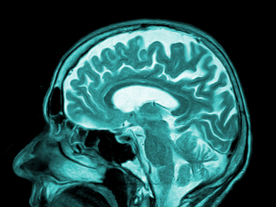
Magnetic resonance imaging (MRI) is based on the phenomenon that the spins of protons in water orientate themselves in a strong external magnetic field. If the magnetic field is switched off, the protons relax back to their native state. This relaxation is measurable and different tissues have characteristic relaxation times (T1) depending on their respective water content. The difference in T1 values is one reason for contrast in MR images. In order to enhance the contrast, paramagnetic metal ions, such as gadolinium(III), are used. High local concentrations of the contrast agent in the body part of interest are key.
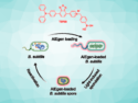
Katharina Landfester, Max Planck Institute of Polymer Research, Mainz, Germany, and colleagues increased the local contrast agent concentration by encapsulation of Gd(III)-complexes (Gadobutrol) in polyurea nanocapsules. Unfortunately, these capsules also restrict the contact between contrast agent and its surroundings, and the positive effect on T1 diminishes.
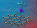
The team found that the proton exchange between surrounding water and the contrast agent capsule is an essential process influencing T1. They tested different aliphatic diamines as polymer building blocks for the capsules and found a high proton exchange rate for diaminobutane (DAB). Gadobutrol encapsulated in DAB leads to an even shorter T1 than an aqueous solution of this contrast agent. A subsequent in vivo study also showed an improved contrast.
- Design and Control of Nanoconfinement to Achieve Magnetic Resonance Contrast Agents with High Relaxivity,
Kerstin Malzahn, Sandro Ebert, Isabel Schlegel, Oliver Neudert, Manfred Wagner, Gunnar Schütz, Andreas Ide, Farnoosh Roohi, Kerstin Münnemann, Daniel Crespy, Katharina Landfester,
Adv. Healthcare Mater. 2015.
DOI: 10.1002/adhm.201500748