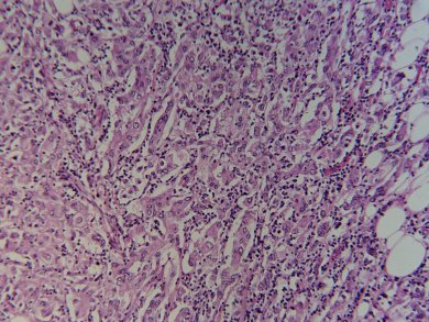
Current diagnostic methods for identification of cancer cells, i.e., stained histopathology, are subjective, based on visual interpretations of cell shape and structure. Stephen Boppart and colleagues, University of Illinois, USA, have developed a quantitative process that produces easy-to-read, color-coded images of cancerous cells in less than five minutes.
By using nonlinear interferometric vibrational imaging (NIVI) to enhance the vibrational frequency of bonds, the team can distinguish between normal cells with high concentrations of lipids and cancerous cells with high protein concentrations. Statistical analysis of the resulting spectrum produces the color-coded image.
In a preclinical rat breast cancer model, NIVI could define cancer boundaries to ±100 μm with greater than 99 % confidence interval, using fresh unstained tissue sections.
- Molecular Histopathology by Spectrally Reconstructed Nonlinear Interferometric Vibrational Imaging
P. D. Chowdary, Z. Jiang, E. J. Chaney, W. A. Benalcazar, D. L. Marks, M. Gruebele, S. A. Boppart,
Cancer Res. 2010, 70.
DOI: 10.1158/0008-547