The visualization of proteins in living cells has been vital to basic biological research. Methods involving labeling with small-molecule dyes are known but are often beset by poor cell permeability of the dye, or off-target binding.
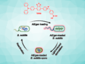
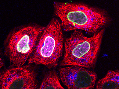
Robert Tampé of Goethe-University, Frankfurt, Germany, and co-workers report a novel method for delivering into living cells a fluorescent small molecule that target proteins of interest carrying a minimal peptide tag. This interaction, between a cell-impermeable nickel complex and a polyhistidine sequence, is well known as the basis of a popular method to purify proteins. The trick involves squeezing the cell under microfluidic control, allowing the dye to rush into the deformed cell before it can repair itself. Once inside the cell, the charged dye cannot leak out and binds with sub-nanomolar affinity to the protein, which is then imaged using super-resolution microscopy.
The researchers demonstrate that this is a general method for labeling a range of proteins in different cell types. Furthermore, the method also worked with a light-activated variant of the nickel complex, allowing imaging with spatio-temporal control.
- Live-Cell Protein Labelling with Nanometre Precision by Cell Squeezing,
A. Kollmannsperger, A. Sharei, A. Raulf, M. Heilemann, R. Langer, K. F. Jensen, R. Wieneke, R. Tampé,
Nat. Commun. 2016.
DOI: 10.1038/ncomms10372