J. Elliott Fowler, Oregon State University, Corvallis, USA, and colleagues have examined why a frog does not lose its insect prey when it retracts its prey-holding tongue forcefully into its mouth. Analysis of frog spit indicated fibrillar structures made of mucin, the main component in mucus, which formed at the moment of pulling. The researchers propose that, in analogy to the fibrils formed by adhesive tape that is peeled off from its substrate, these mucin fibrils may support adhesion between tongue and target.
Protect, Wet, Lubricate, And Glue
Mucus, the slime secreted by mucus glands, is a highly versatile substance. It can protect the most sensitive surfaces of the human body from drying out and from invasion by bacteria and allergens. Nose, lung, mouth, gut, and genitals rely on mucus layers acting as physical barriers, lubricants, and protective coatings. But mucus can also be used for movement. And attachment. This is nowhere more visible than in the snail slime traces traversing your terrace in summer and climbing up the vertical walls of your house and flower boxes.
The main components of mucus are the mucins, glycoproteins with cysteine-rich ends and a core rich in proline, serine, and threonine side chains. With their many hydroxyl functions and polymeric nature, mucins can provide the mucus with a certain adhesiveness. And as mucus covers surfaces, the mucins will be involved in any interaction with the environment.
What Happens at the Tip of the Frog Tongue?
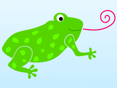
The tongues of frogs are covered with mucus. What happens in it when frogs catch prey? Stanislav Gorb, Kiel University, Germany, who coauthored the paper, has a longstanding interest in adhering mechanisms in the animal kingdom. “The [mucus] liquid itself sticks very well to hydrophilic, but also to hydrophobic surfaces,” he says. However, gluing alone to the hydrophobic and sometimes even superhydrophobic surface of insects is not sufficient. “When the frog retracts the tongue, this liquid must maintain the force,” says Gorb. Otherwise the tongue—and the frog—would lose its prey.
To get insight into the events occurring in the mucus layer, the scientists collected frog mucus just after the tongue hit a substrate. A horned frog in its hunting position was given the sight of a cricket right in front of it. It struck its tongue—only to hit a glass slide placed between the frog and its prey. The tongue mucus left a stain on the glass slide, which the scientists examined using two spectroscopic techniques for surface analysis.
Structural Surface Analysis
Near-edge X-ray absorption fine structure spectroscopy or NEXAFS evaluates unoccupied electron states, thereby indicating the abundance and spatial position of atoms or molecules on a surface. The technology is very sensitive to the bonding environment of the absorbing atom, and it is polarization-dependent, which means that by changing the angle of the incident beam, the orientation of the molecular orbitals relative to the surface can be determined.
The NEXAFS spectra provided evidence for a homogenous composition of the surface mucus. It also indicated the presence of helical structures. Using vibrational spectroscopy, the researchers found directional orientation of the hydrophobic and hydrophilic glycoprotein domains. Helical structure and directional order are indications of fibril formation. The fibrils form as a response to the great pulling force during retraction of the prey-loaded tongue.
Pressure-Sensitive Adhesives
This behavior correlates strikingly with the working principle of pressure-sensitive adhesives, where fibril formation stabilizes adhesion to a substrate during peel-off. Strength and quantity of the fibrils are a measure of debonding ability.
On the frog tongue, mucin fibril formation would instead stabilize prey attachment. Fiber-based adhesion techniques are also known in other places of the animal kingdom. However, in these cases, the fibrils typically do not form inside a liquid or surface layer but are built beforehand as solid- or semisolid structures. Examples are the lamellae architectures of gecko feet or the byssus by which mussels adhere to stones and ship wrecks.
- Surface chemistry of the frog sticky-tongue mechanism,
J. Elliott Fowler, Thomas Kleinteich, Johannes Franz, Cherno Jaye, Daniel A. Fischer, Stanislav N. Gorb, Tobias Weidner, Joe E. Baio,
Biointerphases 2018, 13, 06E408.
https://doi.org/10.1116/1.5052651

